ABG Interpretation Made Simple: The Complete Guide for Nurses
Master ABG interpretation for nurses: the six-step method, all four acid–base disorders, compensation formulas, oxygenation assessment, P/F ratio, mixed disorders, anion gap, clinical patterns, nursing actions, and 26 animated figures — aligned to the 2026 NCLEX-RN® Test Plan.
Quick Answer: What Is the Fastest Way to Interpret an ABG?
Nurses use a six-step systematic method to interpret any arterial blood gas result: look at the pH, assess PaCO₂, assess HCO₃⁻, identify the primary disorder, check for compensation, and evaluate oxygenation. Applying the same steps in the same order every time eliminates omission errors and connects the laboratory result to the nursing action it requires. On the NCLEX-RN®, ABG questions test clinical judgment — the correct answer connects the value pattern to the nursing priority, not to a memorized label alone.
Quick Answer: The nurse's first response to any abnormal ABG is to assess the client, not to interpret the number. Airway, breathing, circulation, mental status, and the oxygen or ventilator setting are evaluated alongside the result. A pH alone does not determine urgency; the client's hemodynamic status, respiratory effort, and trajectory determine the nursing response.
Go deeper: For how ABG findings function as clinical cues inside the CJMM six-step framework, see the clinical judgment guide.
1. Why ABG Interpretation Matters for Nurses
An arterial blood gas, usually called an ABG, provides a rapid picture of three connected areas: ventilation, acid–base balance, and arterial oxygenation. The result can help the health care team understand whether the lungs are removing carbon dioxide, whether the blood is too acidic or too alkaline, and whether oxygen is moving from the lungs into arterial blood.
Entry-level registered nurses working in medical-surgical, critical care, emergency, telemetry, postpartum, or perioperative settings are expected to:
- Recognize ABG patterns that require immediate action versus those that need provider notification within a defined window.
- Connect ABG changes to clinical conditions, respiratory problems, and metabolic disturbances.
- Initiate appropriate nursing interventions based on the client's hemodynamic response.
- Communicate ABG findings using structured handoffs and document the complete clinical sequence.
A strong nurse also asks whether the sample itself is believable. Air in the syringe, delayed analysis, a mislabeled specimen, an incorrect oxygen setting, or a venous sample mistakenly treated as arterial can create misleading values. When a result conflicts sharply with the client's condition, verify the client first and then follow local processes for specimen and analyzer review.
Go deeper: For how the April 2026 NCLEX-RN® Test Plan organizes clinical judgment integration across all Client Needs categories, see the NCLEX-RN® test plan explained.
2. Alignment With the April 2026 NCLEX-RN® Test Plan
The 2026 NCLEX-RN® Test Plan emphasizes application of knowledge, clinical judgment, recognition of trends, diagnostic-test monitoring, laboratory interpretation, and care of clients with impaired ventilation or oxygenation. In Reduction of Risk Potential, entry-level RNs are expected to compare laboratory values with normal values, monitor diagnostic results, obtain specimens, and intervene as needed. In Physiological Adaptation, the plan includes caring for clients on ventilators, managing impaired ventilation or oxygenation, managing fluid and electrolyte imbalances, and responding to acute or life-threatening changes.
Do not study ABGs as four isolated labels. Practise interpreting an unfolding client story: baseline assessment → ABG result → likely primary process → expected compensation → oxygenation → nursing priority → response to intervention.
3. What an ABG Measures
A standard ABG report contains measured values and calculated values. The most commonly used values are pH, PaCO₂, HCO₃⁻, PaO₂, and SaO₂.
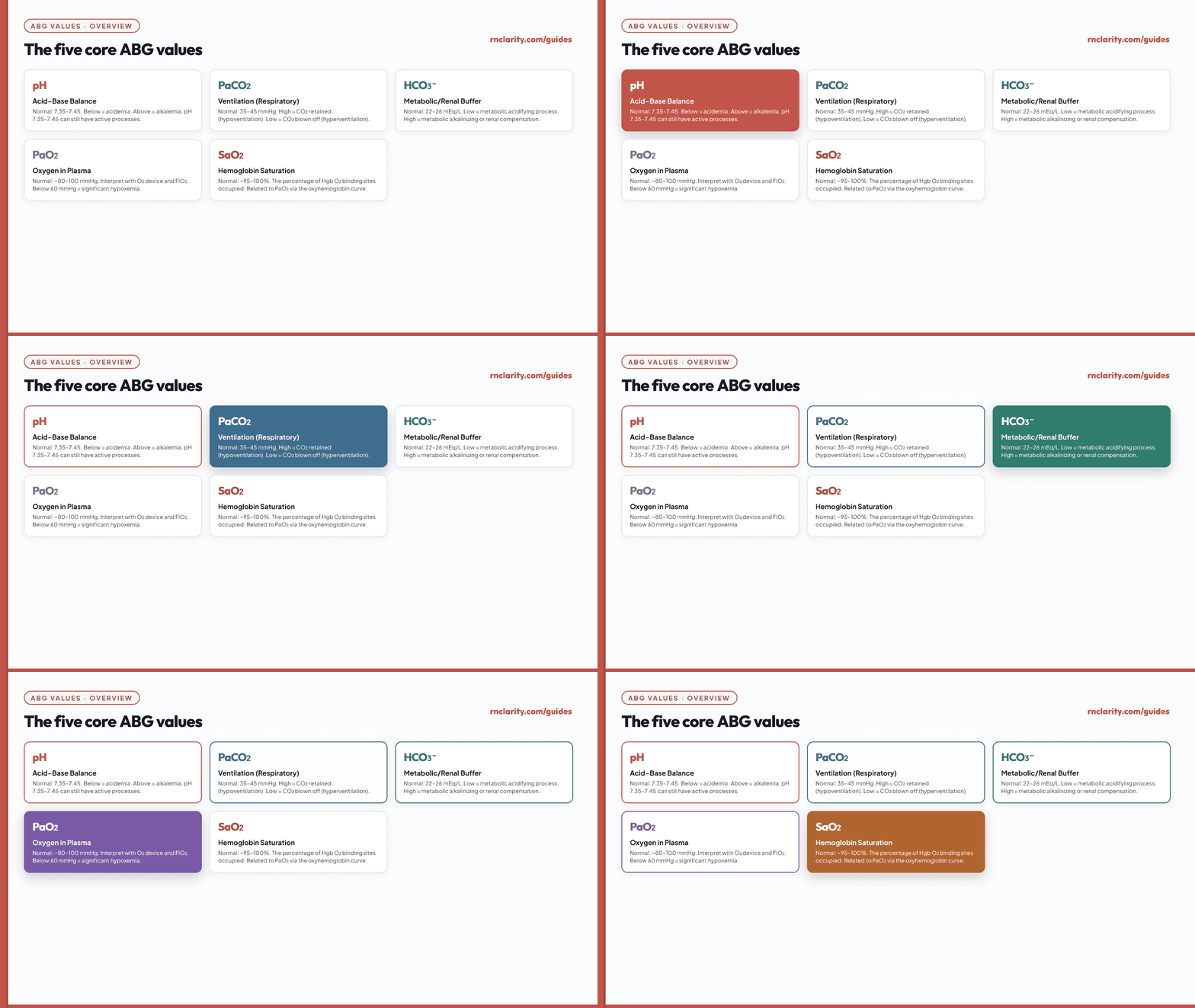
| Value | Common adult teaching range | Primary question |
|---|---|---|
| pH | 7.35–7.45 | Is the blood acidemic, alkalemic, or within the reference range? |
| PaCO₂ | 35–45 mmHg | Is ventilation retaining or removing too much CO₂? |
| HCO₃⁻ | 22–26 mEq/L | Is the metabolic/renal component low, high, or normal? |
| PaO₂ | ~80–100 mmHg at sea level | How much oxygen pressure is dissolved in arterial blood? |
| SaO₂ | ~95–100% | What percentage of hemoglobin sites are oxygenated? |
3.1 pH
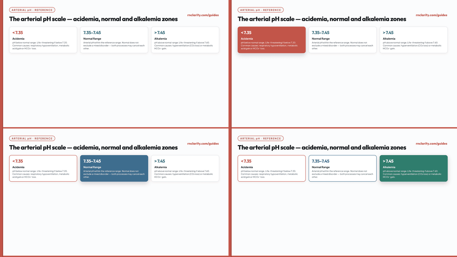
pH describes hydrogen-ion activity and indicates whether arterial blood is acidic or alkaline. A pH below 7.35 is acidemia; a pH above 7.45 is alkalemia. The words acidosis and alkalosis refer to processes that push pH in a direction. A client can have more than one process at the same time, so a normal pH does not prove that acid–base balance is normal.
3.2 PaCO₂
PaCO₂ is the partial pressure of carbon dioxide in arterial blood. It is primarily a ventilation value. Carbon dioxide acts as an acid in the bicarbonate buffer system. When alveolar ventilation decreases, PaCO₂ rises (hypercapnia). When ventilation increases, PaCO₂ falls (hypocapnia). PaCO₂ is not the same as the respiratory rate — a client can breathe rapidly but still ventilate poorly if breaths are shallow.
3.3 HCO₃⁻
Bicarbonate is a major extracellular base. It represents the metabolic or renal side of the interpretation. Low bicarbonate supports a metabolic acidifying process, while high bicarbonate supports a metabolic alkalinizing process or renal compensation for chronic carbon dioxide retention. The HCO₃⁻ on an ABG is often calculated rather than directly measured.
3.4 PaO₂ and SaO₂
PaO₂ is the partial pressure of oxygen dissolved in arterial plasma. SaO₂ is the percentage of hemoglobin oxygen-binding sites occupied by oxygen. They are related through the oxyhemoglobin dissociation curve but are not interchangeable. Oxygen values should never be interpreted without documenting the oxygen device and setting at the time of collection.
4. The Physiology of Acid–Base Balance
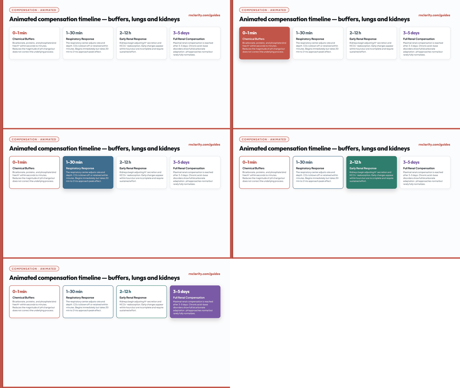
The body produces acids continuously through metabolism. To keep pH within a narrow range, it uses chemical buffers, the lungs, and the kidneys. Buffers act immediately. The lungs can change carbon dioxide within minutes. The kidneys regulate bicarbonate and hydrogen-ion handling over hours to days.
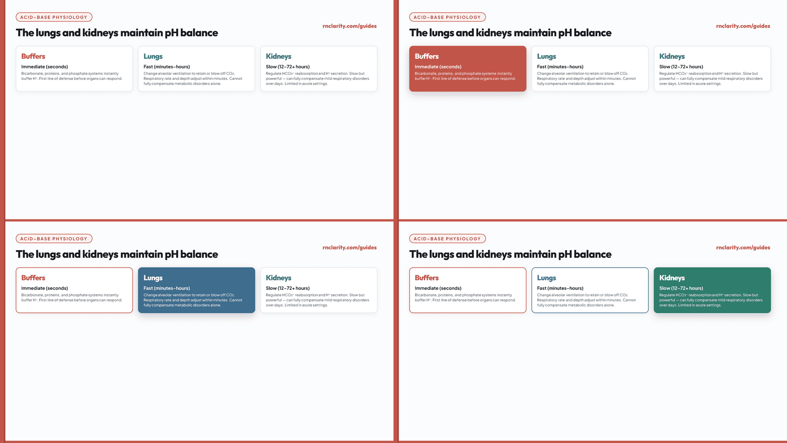
The bicarbonate buffer relationship can be summarized conceptually: pH depends on the balance between bicarbonate and dissolved carbon dioxide. More PaCO₂ pushes toward acidemia. More HCO₃⁻ pushes toward alkalemia. Each primary disorder triggers a compensatory response in the other system to move pH back toward normal.
Go deeper: For how clinical judgment applies to recognizing and prioritizing ABG-related clinical patterns in NGN case studies, see the NGN case studies guide.
5. The Six-Step ABG Interpretation Method
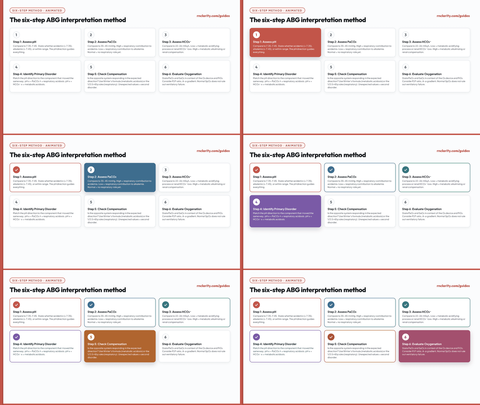
Apply all six steps in sequence for every ABG result.
Step 1 — Assess pH: Compare the measured pH to the reference range (7.35–7.45). State whether it is acidemic, alkalemic, or within range. A borderline pH (7.35–7.37 or 7.43–7.45) may still indicate a significant process; always continue the remaining steps.
Step 2 — Assess PaCO₂: Compare to 35–45 mmHg. A high PaCO₂ is consistent with hypoventilation or CO₂ retention (respiratory contribution to acidemia). A low PaCO₂ is consistent with hyperventilation (respiratory contribution to alkalemia).
Step 3 — Assess HCO₃⁻: Compare to 22–26 mEq/L. A low HCO₃⁻ is consistent with a metabolic acidifying process or renal bicarbonate loss. A high HCO₃⁻ is consistent with a metabolic alkalinizing process or renal compensation for chronic hypercapnia.
Step 4 — Identify the primary disorder: Match the pH direction with the component that moved in the same direction. If pH is acidemic and PaCO₂ is high, the primary disorder is respiratory acidosis. If pH is acidemic and HCO₃⁻ is low, the primary disorder is metabolic acidosis.
Step 5 — Check compensation: Determine whether the opposite system has responded in the expected direction. Fully compensated disorders have near-normal pH but both PaCO₂ and HCO₃⁻ are abnormal. Partial compensation shows movement toward normal pH without reaching it. Use compensation formulas (see Section 10) to judge whether the response is appropriate.
Step 6 — Evaluate oxygenation: State PaO₂ and SaO₂ in the context of the oxygen device and inspired fraction being used. Calculate the P/F ratio or A–a gradient when relevant. A normal PaO₂ on high-flow oxygen is not the same as a normal PaO₂ on room air.
| Step | Assess | Ask |
|---|---|---|
| 1 | pH | Acidemic, alkalemic, or within range? |
| 2 | PaCO₂ | High (retention), low (loss), or normal? |
| 3 | HCO₃⁻ | Low (acidifying), high (alkalinizing), or normal? |
| 4 | Primary disorder | Which component moved with the pH direction? |
| 5 | Compensation | Is the opposite system responding as expected? |
| 6 | Oxygenation | PaO₂ and SaO₂ in context of oxygen delivery? |
6. The Four Primary Acid–Base Disorders
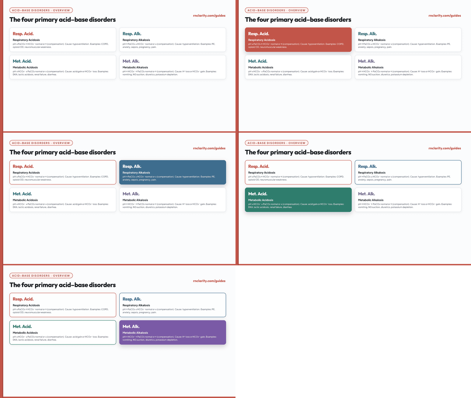
| Disorder | pH | PaCO₂ | HCO₃⁻ | Primary mechanism |
|---|---|---|---|---|
| Respiratory acidosis | ↓ | ↑ | Normal or ↑ (compensation) | Hypoventilation — CO₂ retained |
| Respiratory alkalosis | ↑ | ↓ | Normal or ↓ (compensation) | Hyperventilation — CO₂ blown off |
| Metabolic acidosis | ↓ | Normal or ↓ (compensation) | ↓ | Acid gain or bicarbonate loss |
| Metabolic alkalosis | ↑ | Normal or ↑ (compensation) | ↑ | Hydrogen-ion loss or bicarbonate gain |
7. Respiratory Acidosis
Respiratory acidosis begins with inadequate alveolar ventilation that allows carbon dioxide to accumulate. PaCO₂ rises and pH falls. The kidneys respond over hours to days by retaining bicarbonate.
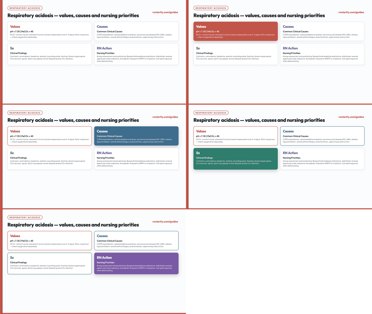
Common causes:
- COPD exacerbation with air trapping and reduced alveolar ventilation.
- Opioid, benzodiazepine, or sedative-induced respiratory depression.
- Neuromuscular disorders (myasthenia gravis, Guillain-Barré, ALS) that reduce respiratory muscle strength.
- Obesity hypoventilation syndrome.
- Severe asthma or status asthmaticus with respiratory fatigue.
- Pneumothorax, pleural effusion, or pneumonia impairing gas exchange.
- Upper airway obstruction or laryngospasm.
Clinical findings may include confusion, somnolence, headache, asterixis, flushing, bounding pulse, and respiratory fatigue. Severe hypercapnia (PaCO₂ > 70–80 mmHg acutely) can cause CO₂ narcosis.
Nursing priorities: Assess airway and maintain patency. Position for optimal breathing (semi-Fowler's or high-Fowler's when tolerated). Support ventilation according to orders and protocols. Check the oxygen delivery system. Review sedating medications and administer a prescribed reversal agent when indicated (naloxone for opioids, flumazenil for benzodiazepines when authorized). Prepare for noninvasive or invasive ventilatory support. Reassess after any intervention.
NCLEX-RN® caution: A normal or high oxygen saturation does not prove that ventilation is adequate. A client can retain a dangerous amount of carbon dioxide while receiving supplemental oxygen.
8. Respiratory Alkalosis
Respiratory alkalosis begins with ventilation that removes carbon dioxide faster than the body produces it. PaCO₂ falls and pH rises. In a sustained process, renal bicarbonate decreases. The nurse must determine why the client is hyperventilating rather than assuming anxiety.
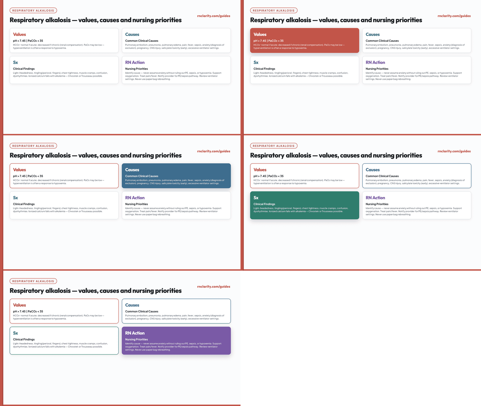
Common causes:
- Hypoxemia from pulmonary embolism, pneumonia, pulmonary edema, or high altitude.
- Pain, fever, or anxiety — only after urgent physiologic causes have been assessed.
- Sepsis and systemic inflammatory states.
- Pregnancy-related physiologic hyperventilation.
- Central nervous system stimulation or injury.
- Early salicylate toxicity.
- Excessive minute ventilation on a mechanical ventilator.
Clinical findings may include light-headedness, tingling around the mouth or fingers, chest discomfort, muscle cramps, confusion, or dysrhythmias from changes in ionized calcium and cerebral blood flow.
Nursing actions: Support oxygenation when indicated. Treat pain or fever according to orders. Reduce environmental triggers when anxiety is likely after physiologic causes are excluded. Notify the appropriate clinician about possible pulmonary embolism or sepsis. Review ventilator settings with the respiratory care and prescribing team.
NCLEX-RN® trap: Routine rebreathing into a paper bag is unsafe because it can worsen hypoxemia and delay recognition of a serious cause of hyperventilation.
Go deeper: For how to connect respiratory findings to clinical judgment priorities in practice questions, see the practice questions guide.
9. Metabolic Acidosis
Metabolic acidosis begins with loss of bicarbonate, accumulation of nonvolatile acid, or impaired renal acid excretion. HCO₃⁻ is low and pH is low or pushed downward. The lungs usually compensate by lowering PaCO₂ through increased ventilation. The breathing pattern may become deep and rapid when acidosis is severe — this is called Kussmaul respiration.
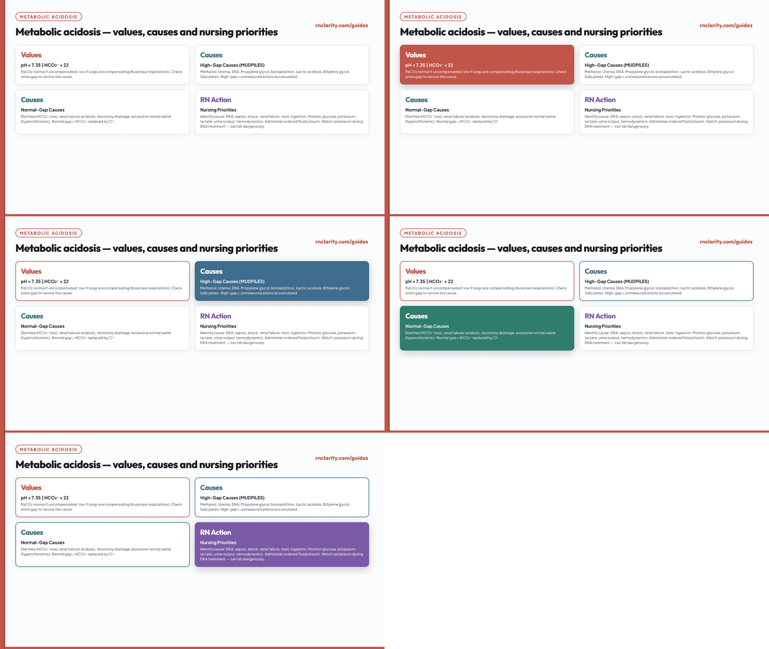
Common causes:
- Ketoacidosis from diabetes (DKA), starvation, or alcohol.
- Lactic acidosis from tissue hypoperfusion, shock, severe sepsis, or seizures.
- Kidney failure or renal tubular disorders that impair acid excretion or bicarbonate handling.
- Bicarbonate loss from severe diarrhea, intestinal drainage, or selected fistulas.
- Toxic ingestions and medication-related causes (methanol, ethylene glycol, salicylates).
Nursing priorities: Identify shock, sepsis, DKA, renal failure, or toxic exposure. Monitor hemodynamics, mental status, glucose, electrolytes, and urine output. Support airway and breathing. Administer ordered fluids, insulin, or other therapies. Evaluate trends — a falling potassium during DKA treatment can become immediately dangerous even while pH improves.
10. Metabolic Alkalosis
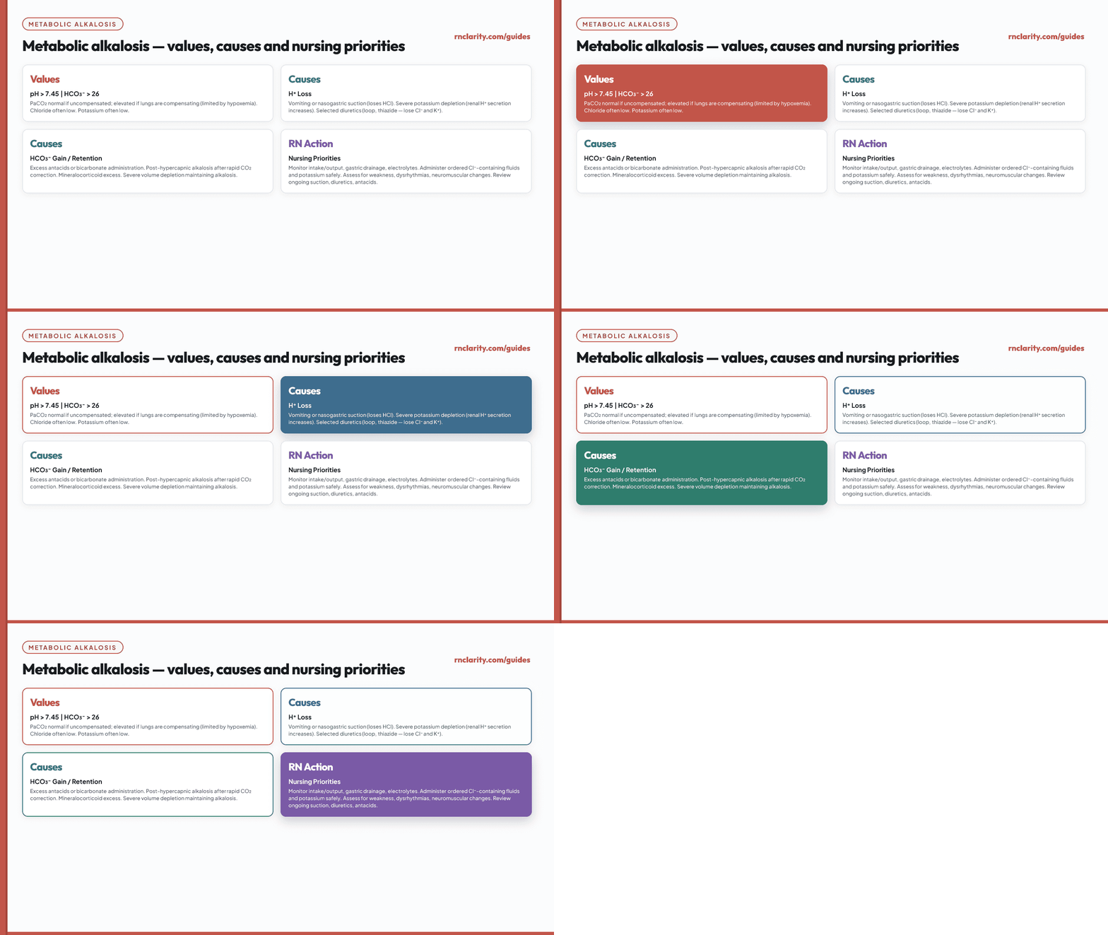
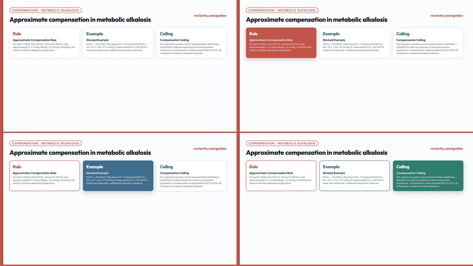
Metabolic alkalosis begins with hydrogen-ion loss, bicarbonate gain, or maintenance of alkalosis through volume, chloride, or potassium disturbances. HCO₃⁻ is high and pH is high or pushed upward. PaCO₂ may rise through compensatory hypoventilation, but the response is limited by oxygen needs.
Common causes:
- Vomiting or gastric suction with loss of hydrochloric acid.
- Diuretic-related volume, chloride, and potassium loss.
- Excess alkali administration in susceptible clients.
- Post-hypercapnic alkalosis after rapid correction of chronic carbon dioxide retention.
- Severe potassium depletion.
Nursing actions: Monitor intake and output, gastric drainage, hemodynamics, and electrolytes. Administer ordered chloride-containing fluids or potassium safely. Evaluate the need for ongoing suction or diuretic therapy with the team. Watch for dysrhythmias or neuromuscular changes (weakness, cramps, paresthesias).
11. Compensation and Expected Responses
Compensation rules answer a focused question: Is the second system responding approximately as expected to the primary disorder? They are most useful after the primary process has been identified.
11.1 Metabolic Acidosis: Winter's Formula
For a primary metabolic acidosis, expected respiratory compensation is estimated with Winter's formula:
Expected PaCO₂ = 1.5 × HCO₃⁻ + 8 (± 2 mmHg)
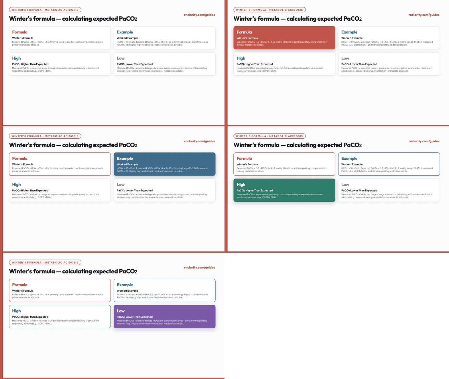
Example: pH 7.20, PaCO₂ 26 mmHg, HCO₃⁻ 10 mEq/L. Expected PaCO₂ = 1.5 × 10 + 8 = 23 ± 2, or about 21–25 mmHg. Measured 26 is close but slightly above. If the measured PaCO₂ is higher than expected, an additional respiratory acidosis is present. If lower, an additional respiratory alkalosis is present.
11.2 Metabolic Alkalosis
A commonly taught estimate is that PaCO₂ rises by about 0.7 mmHg for each 1 mEq/L rise in HCO₃⁻ above 24.
11.3 Respiratory Acidosis
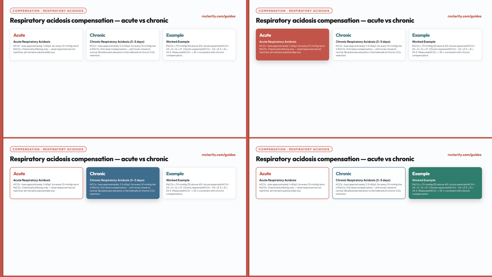
For acute respiratory acidosis, HCO₃⁻ rises approximately 1 mEq/L for every 10 mmHg rise in PaCO₂. For chronic respiratory acidosis (after 3–5 days of renal compensation), HCO₃⁻ rises approximately 3.5 mEq/L for every 10 mmHg rise in PaCO₂.
11.4 Respiratory Alkalosis
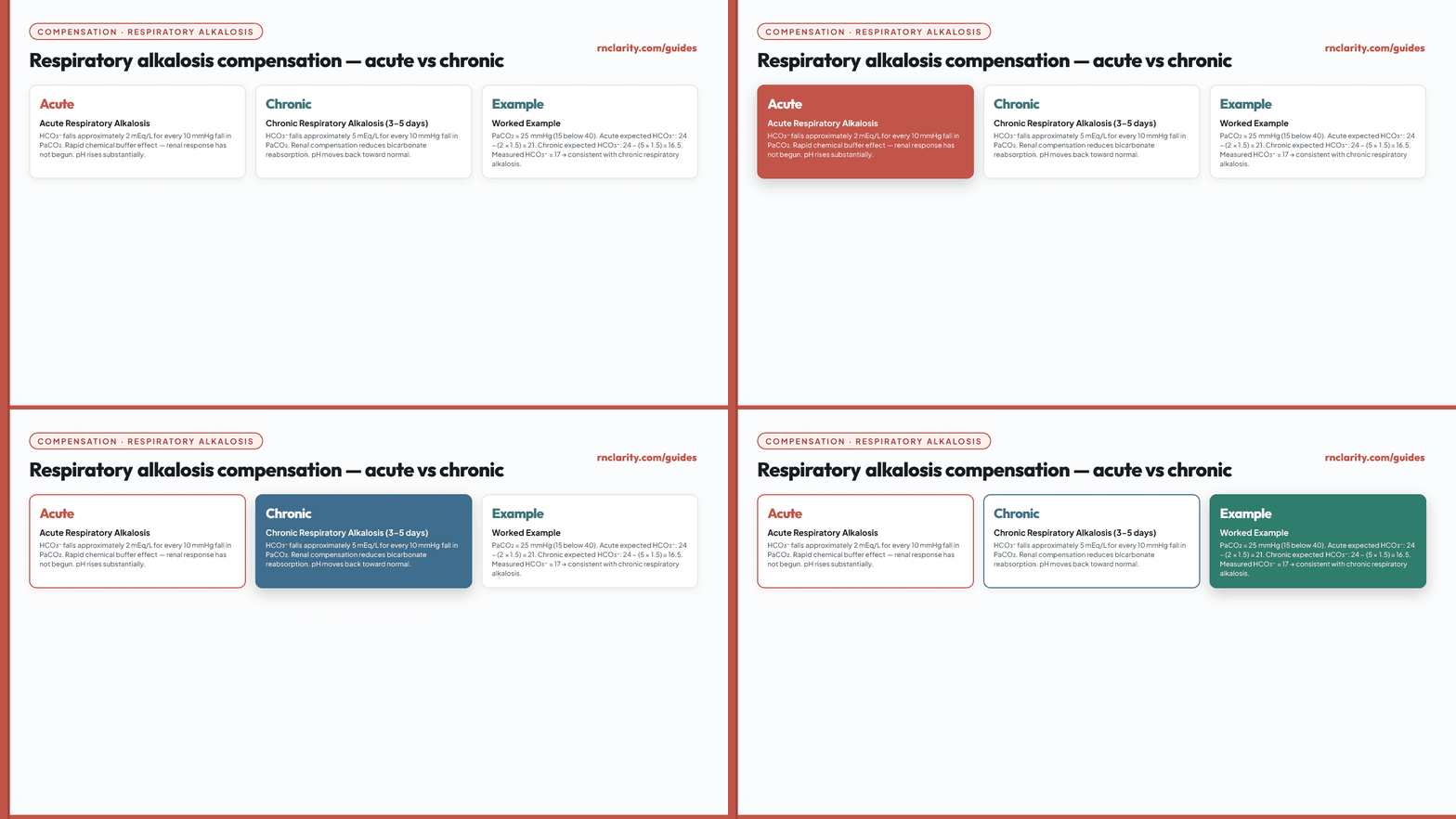
For acute respiratory alkalosis, HCO₃⁻ falls approximately 2 mEq/L for every 10 mmHg fall in PaCO₂. For chronic respiratory alkalosis, HCO₃⁻ falls approximately 5 mEq/L per 10 mmHg.
12. Mixed Acid–Base Disorders
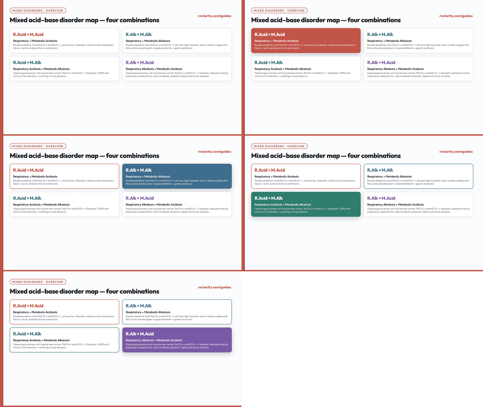
A mixed disorder occurs when more than one primary acid–base process is present simultaneously. Unlike simple disorders with compensation, both PaCO₂ and HCO₃⁻ may be abnormal in the same direction, both normal despite an abnormal pH, or pH may be normal while both components are strikingly abnormal.
Common clinical examples of mixed disorders:
| Clinical setting | Mix | Key clue |
|---|---|---|
| Cardiac arrest or severe shock | Respiratory acidosis + metabolic acidosis | Both pH very low; PaCO₂ high and HCO₃⁻ low simultaneously |
| Vomiting + severe COPD | Metabolic alkalosis + respiratory acidosis | pH may be near normal despite both components abnormal |
| Salicylate toxicity (early) | Respiratory alkalosis + metabolic acidosis | pH may be near normal; PaCO₂ low and HCO₃⁻ low |
| Post-cardiac surgery with NG suction | Metabolic alkalosis + respiratory alkalosis | pH very high; both PaCO₂ low and HCO₃⁻ high |
To identify a mixed disorder, use compensation formulas. When the measured component differs substantially from the expected compensation range, a second primary disorder is likely.
Go deeper: For how mixed ABG patterns connect to clinical judgment priorities in the NCLEX-RN® practice question bank, see the practice questions guide.
13. Anion Gap and Advanced Metabolic Interpretation
The anion gap (AG) is a calculated value that helps organize the causes of metabolic acidosis. It estimates the concentration of unmeasured anions in the plasma.
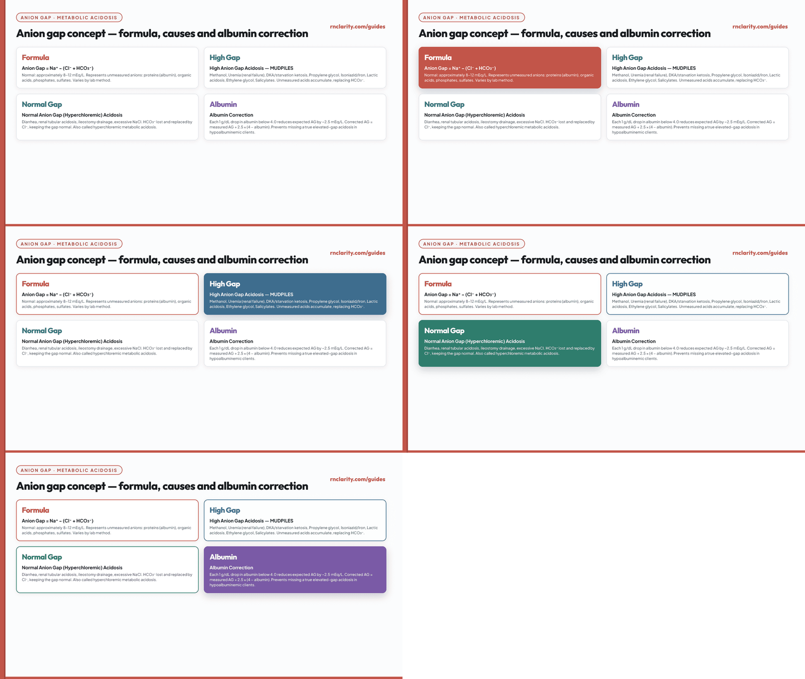
Anion gap formula: AG = Na⁺ − (Cl⁻ + HCO₃⁻). Common adult reference range: approximately 8–12 mEq/L (varies by laboratory).
High anion gap metabolic acidosis (MUDPILES): Methanol, Uremia, DKA, Propylene glycol, Isoniazid/Iron, Lactic acidosis, Ethylene glycol, Salicylates.
Normal anion gap (hyperchloremic) metabolic acidosis: Diarrhea (bicarbonate loss), renal tubular acidosis, certain medications, excessive normal saline, ileostomy or intestinal drainage.
Albumin correction: Each 1 g/dL decrease in serum albumin below 4.0 g/dL decreases the expected anion gap by approximately 2.5 mEq/L. In hypoalbuminemic clients, the corrected anion gap should be used to avoid missing a true elevated gap acidosis.
14. Oxygenation: PaO₂, SaO₂, SpO₂, and the Oxyhemoglobin Dissociation Curve
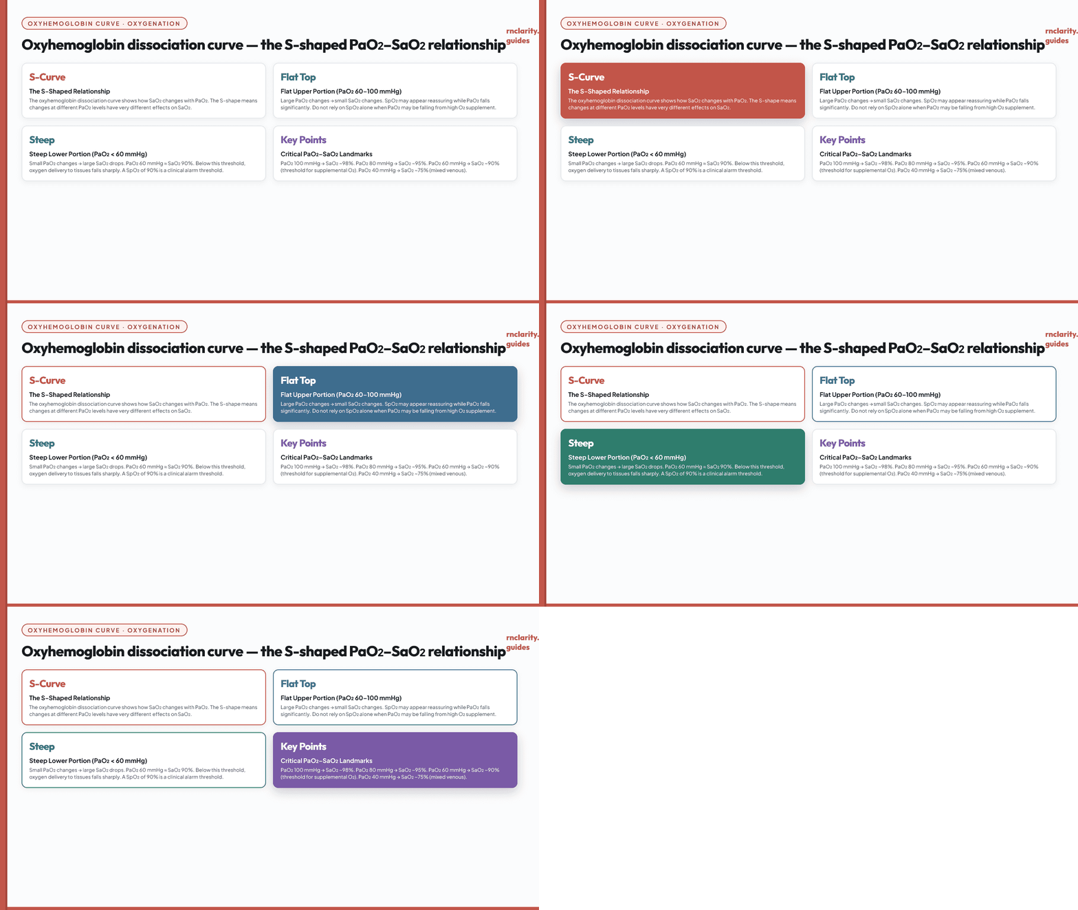
The oxyhemoglobin dissociation curve shows the S-shaped relationship between PaO₂ (the driving pressure of oxygen dissolved in plasma) and SaO₂ (the percentage of hemoglobin bound to oxygen). The curve has two clinically important features:
- The flat upper portion (PaO₂ 70–100 mmHg): A large drop in PaO₂ produces only a small drop in SaO₂. This means pulse oximetry may appear reassuring while PaO₂ is falling significantly.
- The steep lower portion (PaO₂ < 60 mmHg): A small drop in PaO₂ produces a large drop in SaO₂. When SpO₂ falls below about 90%, oxygen delivery to tissues drops sharply.
14.1 Curve Shifts
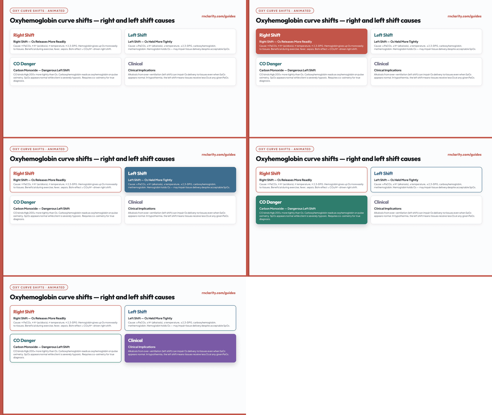
Right shift (hemoglobin releases oxygen more readily — beneficial for tissues during increased demand):
- Increased PaCO₂ (Bohr effect)
- Acidemia (lower pH)
- Increased 2,3-DPG
- Increased temperature
Left shift (hemoglobin holds oxygen more tightly — may impair tissue delivery despite acceptable SpO₂):
- Decreased PaCO₂
- Alkalemia (higher pH)
- Decreased 2,3-DPG (stored blood)
- Decreased temperature, carbon monoxide poisoning, methemoglobinemia
14.2 PaO₂–SaO₂ Relationship
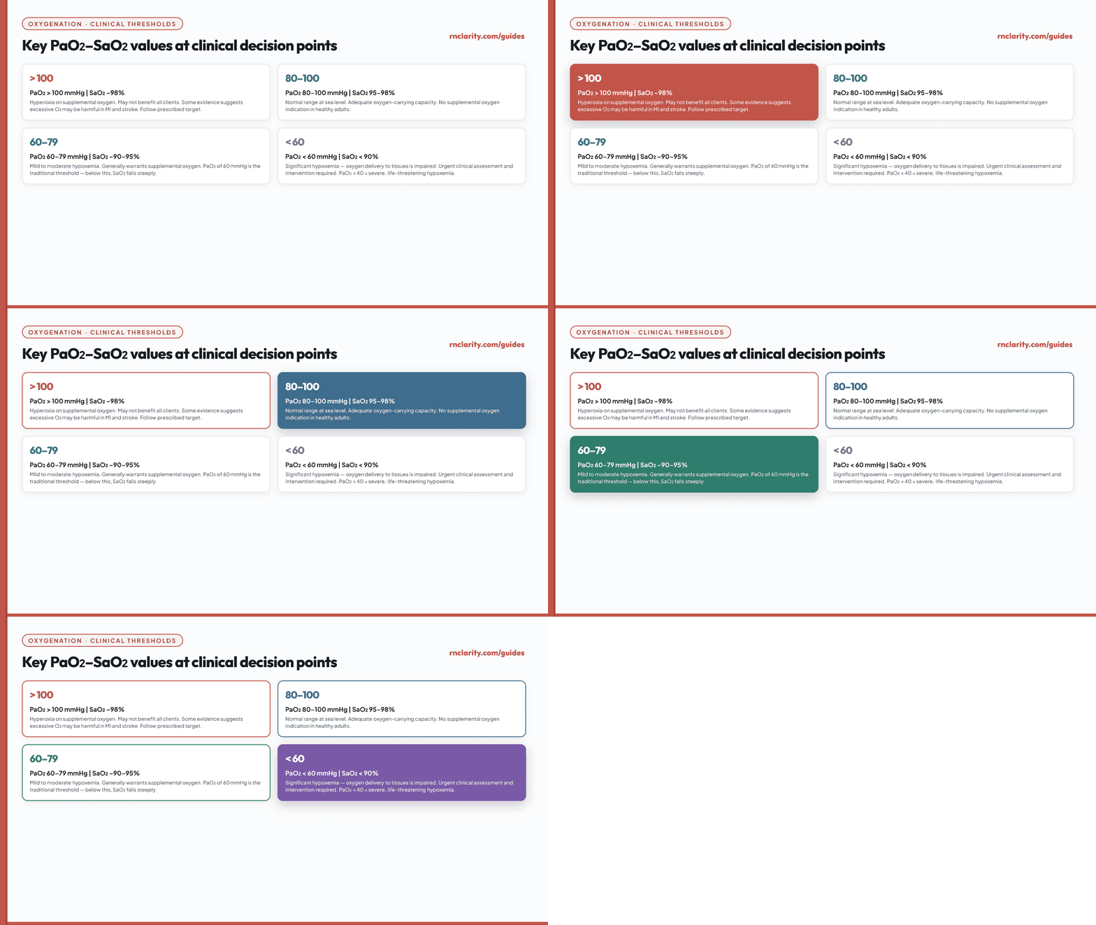
Clinical significance of PaO₂ thresholds:
| PaO₂ | SaO₂ (approximate) | Clinical significance |
|---|---|---|
| > 100 mmHg | ~98% | Hyperoxia on supplemental O₂; may not be beneficial for all clients |
| 80–100 mmHg | 95–98% | Normal range at sea level |
| 60–79 mmHg | ~90–95% | Mild to moderate hypoxemia; usually warrants supplemental oxygen |
| 40–59 mmHg | ~75–89% | Moderate to severe hypoxemia; urgent intervention needed |
| < 40 mmHg | < 75% | Severe hypoxemia; life-threatening |
15. P/F Ratio and A–a Gradient
15.1 P/F Ratio
The P/F ratio (PaO₂/FiO₂) standardizes oxygenation for the fraction of inspired oxygen being delivered. It allows comparison of oxygenation across different oxygen settings.
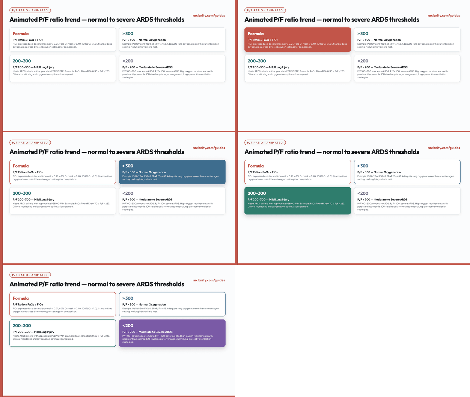
P/F ratio = PaO₂ ÷ FiO₂ (FiO₂ expressed as a decimal: room air = 0.21; 100% O₂ = 1.00)
| P/F Ratio | Interpretation |
|---|---|
| > 300 | Normal oxygenation |
| 200–300 | Mild lung injury (ARDS criteria) |
| 100–200 | Moderate ARDS |
| < 100 | Severe ARDS |
15.2 A–a Gradient
The alveolar-arterial (A–a) gradient compares the oxygen partial pressure calculated in the alveoli with the measured PaO₂ in arterial blood. A widened A–a gradient suggests a problem between the alveolus and the arterial blood (V/Q mismatch, diffusion impairment, or shunt).
![Animated A-a gradient concept showing the alveolar gas equation: PAO2 = (FiO2 x [Patm - PH2O]) - (PaCO2 / 0.8). A-a gradient = PAO2 - PaO2. Normal A-a gradient on room air is approximately 5-15 mmHg, increasing with age. A widened gradient suggests V/Q mismatch, shunt, or diffusion impairment.](/guides/abg-interpretation-for-nurses/fig21-aa-gradient-concept.gif)
Normal A–a gradient (approximate, room air): 5–15 mmHg in young adults; increases with age and inspired oxygen fraction.
Causes of widened A–a gradient: Pulmonary embolism, pneumonia, ARDS, pulmonary edema, atelectasis, intracardiac shunt.
Normal A–a gradient with hypoxemia: Suggests hypoventilation as the cause of low PaO₂ (e.g., opioid-induced, neuromuscular).
16. ABG Collection, Safety, and Specimen Errors
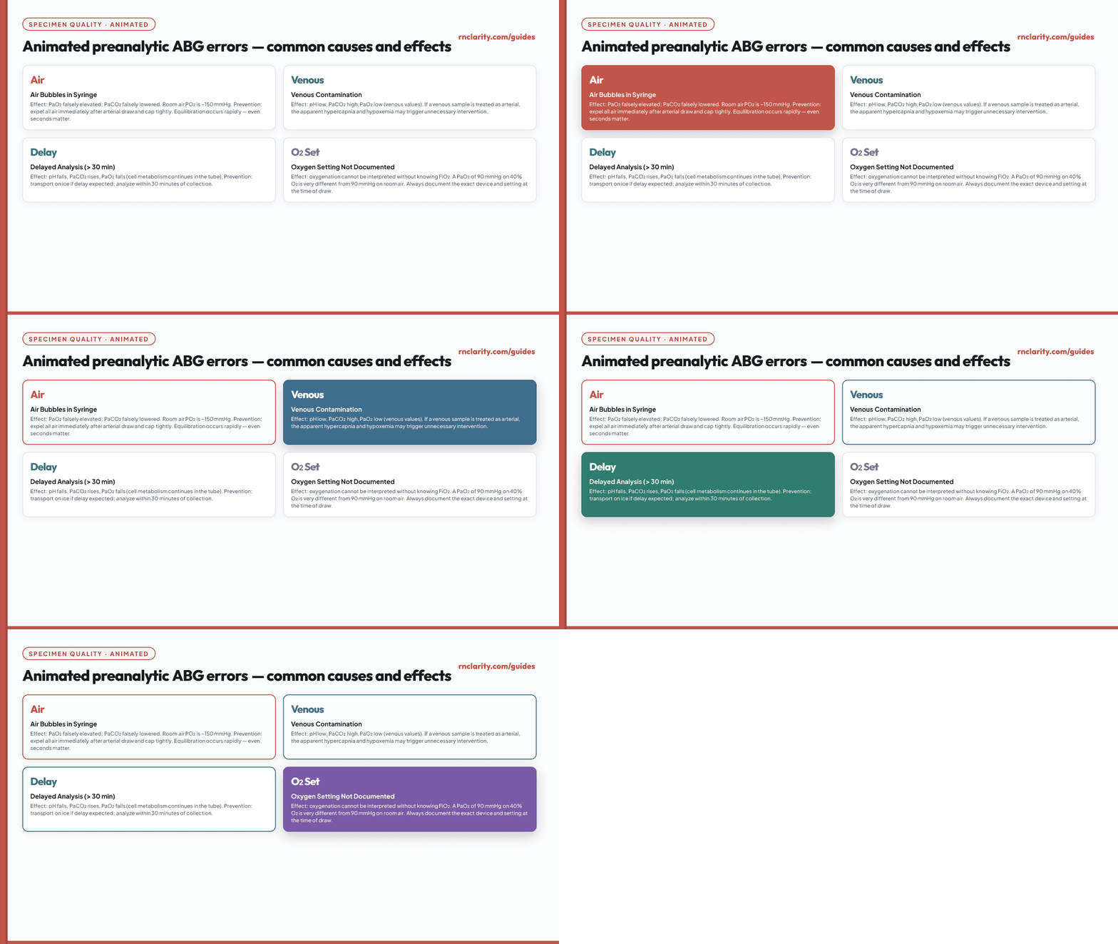
ABG specimens are fragile and time-sensitive. Pre-analytic errors can produce misleading values that lead to incorrect clinical decisions.
| Error | Effect on result | Nursing prevention |
|---|---|---|
| Air in syringe | PaO₂ falsely elevated; PaCO₂ falsely lowered | Expel all air immediately after collection; cap tightly |
| Venous contamination | Low pH, high PaCO₂, low PaO₂ | Confirm arterial waveform before drawing; use proper technique |
| Delayed analysis (> 30 min) | pH falls, PaCO₂ rises, PaO₂ falls | Transport on ice if delay expected; analyze within 30 min |
| Incorrect oxygen setting | Oxygenation misinterpreted | Document exact FiO₂ or oxygen device at time of draw |
| Mislabeled specimen | Wrong patient action | Apply two-identifier verification before labeling |
| Excess heparin in syringe | Dilutional effect on all values | Use pre-heparinized syringes; do not overfill |
When a result conflicts sharply with the client's clinical picture, the nurse should assess the client, consider pre-analytic error, and follow local policy for specimen repeat before acting on a potentially erroneous value — unless the client is in immediate danger.
17. ABG Versus VBG and Pulse Oximetry
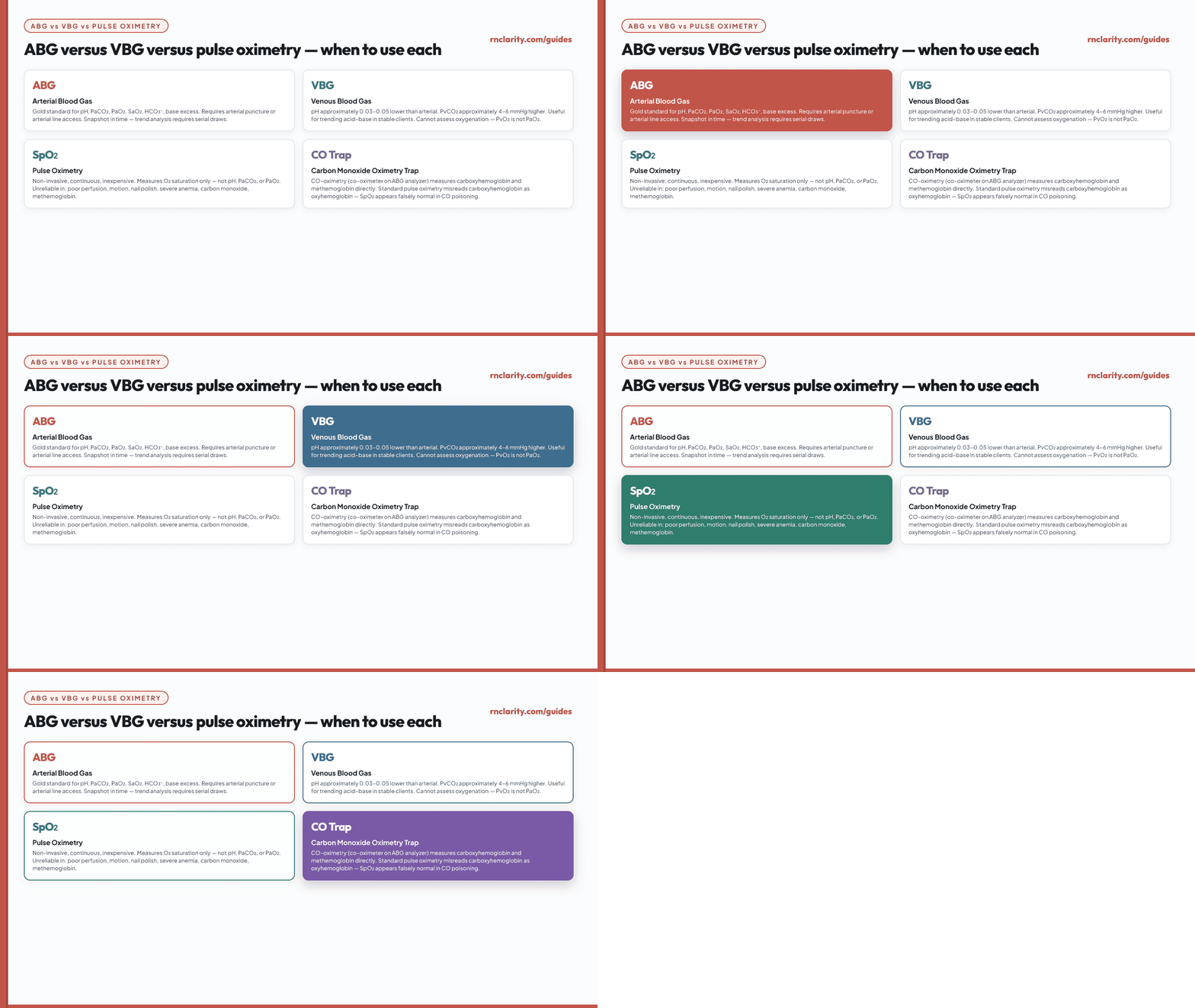
| Measure | Source | Strengths | Limitations |
|---|---|---|---|
| ABG | Arterial puncture or line | Gold standard for pH, PaCO₂, PaO₂, SaO₂, HCO₃⁻ | Invasive; snapshot in time; operator-dependent |
| VBG | Venous blood | Easier to obtain; pH correlates reasonably in stable clients (approximately 0.03–0.05 lower); PvCO₂ approximately 4–6 mmHg higher | Cannot reliably assess oxygenation (PvO₂ is not PaO₂) |
| SpO₂ | Pulse oximetry | Continuous; non-invasive | Affected by perfusion, motion, pigmentation, nail polish, carbon monoxide, methemoglobin |
SpO₂ limitations that matter clinically:
- Carbon monoxide poisoning: carboxyhemoglobin reads as oxyhemoglobin — SpO₂ appears falsely normal.
- Methemoglobinemia: SpO₂ drifts toward approximately 85% regardless of severity.
- Severe anemia: SpO₂ may be normal while total oxygen-carrying capacity is critically low.
- Shock or poor perfusion: SpO₂ reading is unreliable when signal is poor.
18. ABGs in Mechanical Ventilation and Oxygen Therapy
ABGs are essential for managing mechanically ventilated clients because they confirm that ventilator settings are achieving the intended physiologic goals. The respiratory care therapist and prescribing clinician interpret the ventilator-related components, but the bedside nurse recognizes concerning changes and escalates appropriately.
Key nursing responsibilities for ventilated clients:
- Document the ventilator settings (FiO₂, mode, tidal volume, PEEP, rate) at the time of ABG collection.
- Assess for patient–ventilator dyssynchrony: agitation, paradoxical breathing, high-pressure alarms, desaturation.
- Monitor for complications: barotrauma, volutrauma, inadvertent PEEP, secretion accumulation.
- Ensure head-of-bed elevation at 30–45 degrees unless contraindicated (ventilator-associated pneumonia prevention).
- Never adjust ventilator settings without an authorized order except in emergency rescue situations per protocol.
Oxygen therapy targets: Oxygen targets are individualized. For a person with COPD or another risk factor for hypercapnic respiratory failure, an initial target of 88–92% is commonly used during an acute illness while blood gases are obtained; the target may change after the results and clinical history are reviewed. Controlled oxygen aims to avoid both hypoxemia and excess oxygen. Oxygen-induced CO₂ retention has several mechanisms and should not be explained solely as loss of “hypoxic drive.” Follow the prescribed target, reassess the patient, and escalate deterioration.
19. Common Clinical Patterns
Go deeper: For how laboratory findings connect to clinical judgment priorities across body systems in the NCLEX-RN® study plan, see the study plan guide.
19.1 Diabetic Ketoacidosis (DKA)
Pattern: Metabolic acidosis with high anion gap. Low HCO₃⁻, low pH. PaCO₂ low (Kussmaul compensation). Glucose elevated. Ketones positive. Potassium may be high initially then drops rapidly with insulin treatment.
Nursing priorities: Fluid resuscitation, insulin infusion per protocol, potassium monitoring (critical — can drop to dangerous levels during treatment), glucose monitoring, anion gap trending, mental status assessment.
19.2 COPD Exacerbation
Pattern: Respiratory acidosis, often chronic (elevated baseline bicarbonate). pH lower than baseline. PaCO₂ higher than baseline. Oxygenation may be impaired. Consider acute-on-chronic hypercapnia when the pH is more acidemic than expected for the degree of bicarbonate elevation.
Nursing priorities: Titrate oxygen to the prescribed target, obtain or review blood gases, give bronchodilators per order, position for easier breathing, assess mental status and respiratory effort, and prepare for noninvasive ventilation when indicated. An initial 88–92% target may be used during an acute COPD exacerbation pending blood gases; COPD alone does not determine one target for every patient.
19.3 Pulmonary Embolism
Pattern: Respiratory alkalosis and hypoxemia are possible but not diagnostic. PaCO₂ may be low. PaO₂ may be low. A–a gradient may be widened. A normal ABG does not exclude pulmonary embolism.
19.4 Sepsis
Pattern: Early sepsis may show respiratory alkalosis from compensatory hyperventilation. As perfusion falls, lactic acidosis and metabolic acidosis develop. Mixed respiratory alkalosis and metabolic acidosis is common.
19.5 Renal Failure
Pattern: Metabolic acidosis with low to normal HCO₃⁻. Potassium may be elevated. Oliguria, edema, uremic symptoms.
Nursing priorities: Monitor potassium, ECG, fluid status, and dialysis plan. Report urgent changes promptly.
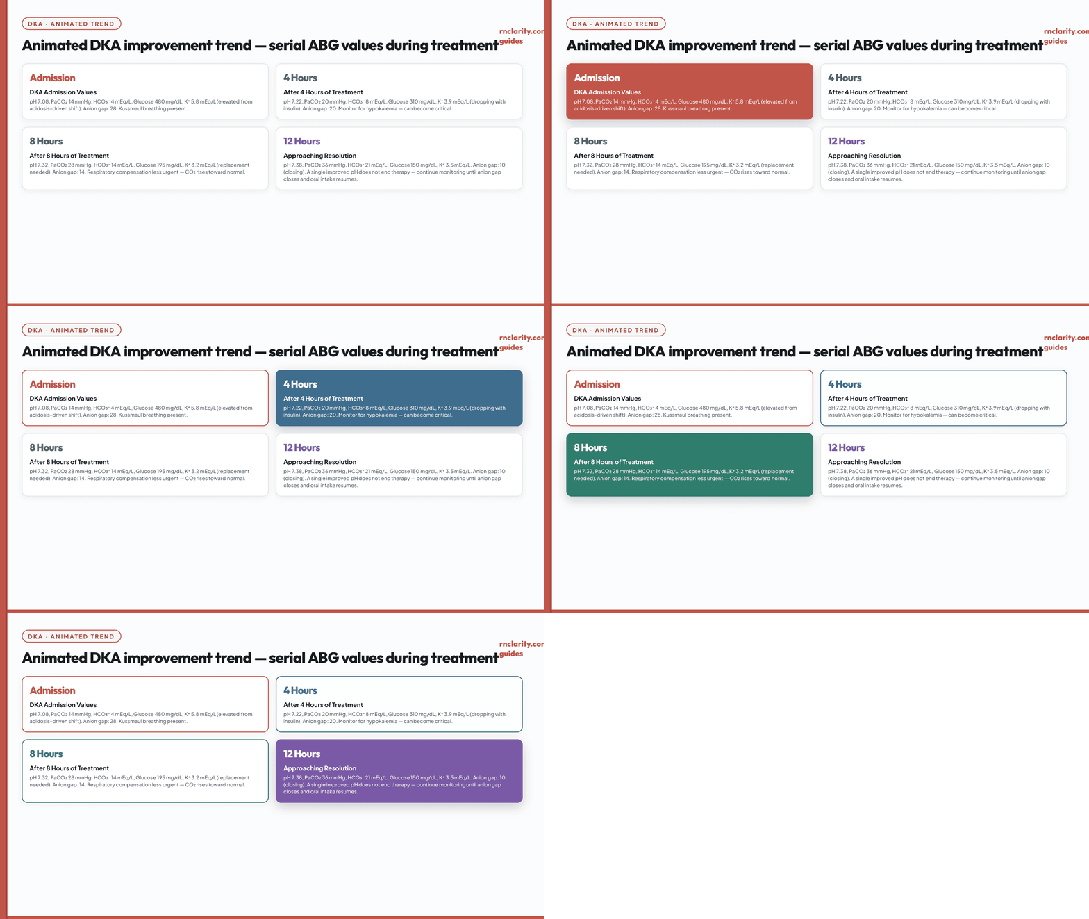
In DKA, improvement commonly appears as rising bicarbonate and pH with PaCO₂ moving upward toward normal. The nurse continues monitoring potassium, glucose, anion gap, neurologic status, fluid balance, and treatment complications. A single improved pH does not mean therapy is complete.
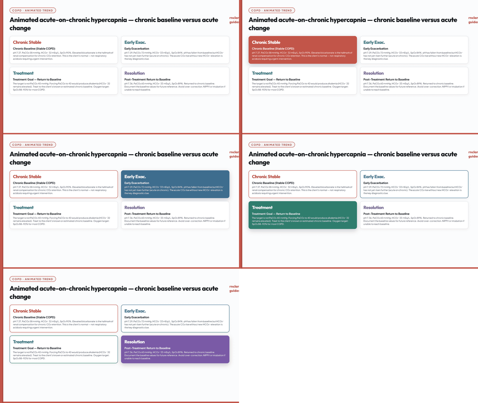
In chronic carbon dioxide retention, the goal is not necessarily a PaCO₂ of 40 mmHg. Rapidly forcing PaCO₂ to normal can produce alkalemia in chronically hypercapnic clients whose renal compensation has elevated bicarbonate. Treatment targets are individualized by the clinical team.
20. Nursing Actions and Escalation
Nursing action is guided by the client's stability, not by a universal ABG number. Many laboratories have critical-result thresholds, but those thresholds vary. A pH near 7.20 may be expected temporarily during a controlled treatment plan in one setting and an immediate emergency in another. The nurse follows the institution's critical-value policy while using clinical judgment.
When to escalate urgently:
- New or worsening altered mental status, apnea, severe respiratory fatigue, or inability to protect the airway.
- Rapidly falling oxygen saturation, severe hypoxemia, or increasing oxygen requirement.
- Acidemia or alkalemia accompanied by hemodynamic instability, dysrhythmia, seizure, or neurologic change.
- Unexpected PaCO₂ rise with reduced ventilation or a deteriorating ventilated client.
- Suspected DKA, severe sepsis, toxic ingestion, renal failure with hyperkalemia, or other life-threatening metabolic cause.
SBAR example for ABG escalation: "The client is increasingly drowsy after opioids. Respiratory rate is 7 with shallow breaths on 2 L nasal cannula. ABG is pH 7.19, PaCO₂ 72, HCO₃⁻ 27, PaO₂ 84. I am supporting the airway, have called rapid response, and am requesting immediate evaluation for opioid-induced ventilatory failure."
Go deeper: For how to integrate clinical judgment into NCLEX-RN® exam strategy for ABG and laboratory questions, see the practice questions guide.
21. NCLEX-RN® Clinical Judgment and ABG Questions
ABG questions on the NCLEX-RN® may appear as stand-alone items or as part of an unfolding clinical judgment case. The examination tests what the nurse should recognize, prioritize, do, or evaluate — not a memorized label alone.
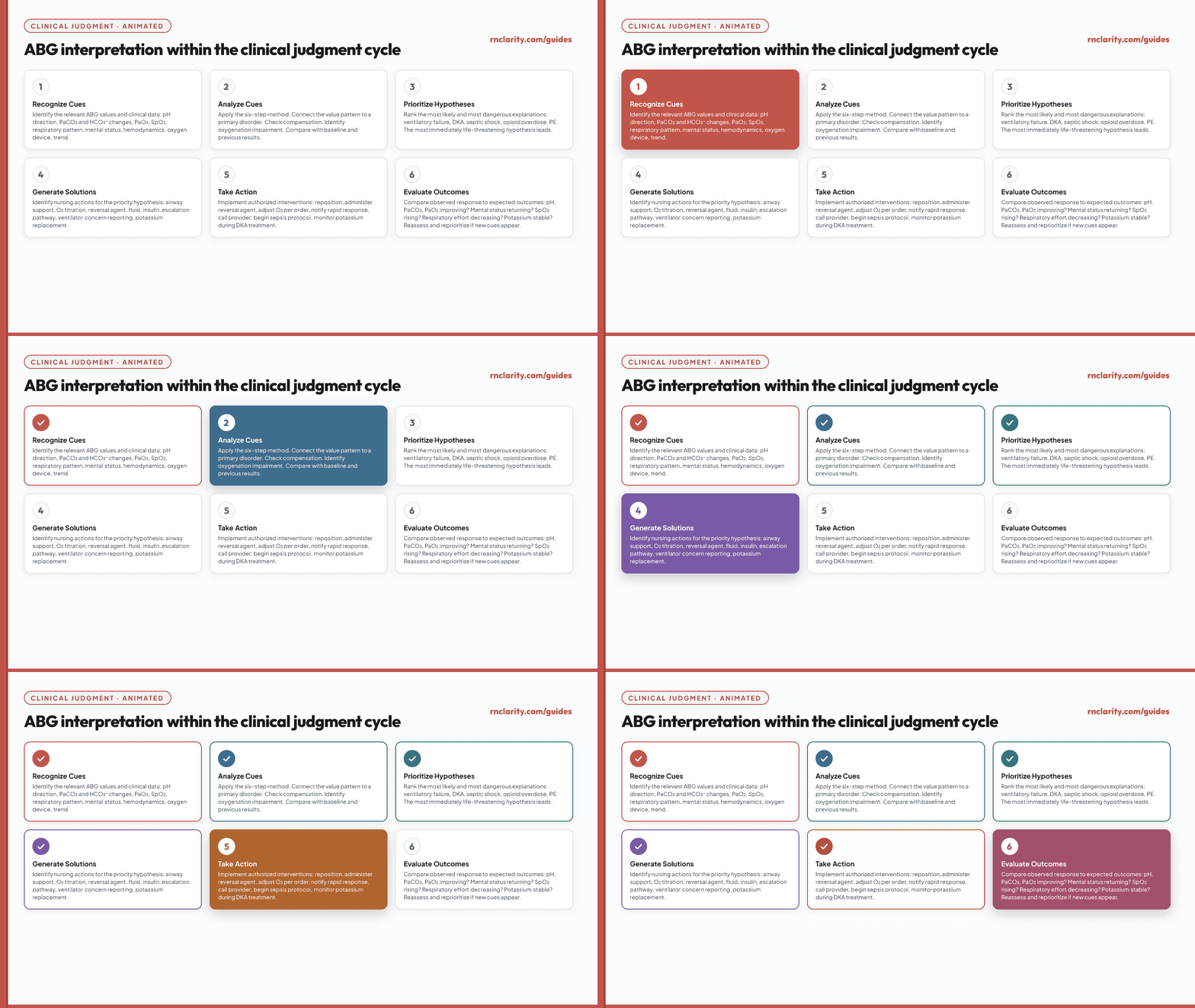
Six-step clinical judgment framework applied to ABG items:
Recognize cues: Identify the ABG values that are clinically relevant, but also select assessment cues that support the interpretation: declining mental status with high PaCO₂, Kussmaul breathing with low HCO₃⁻, worsening oxygenation despite higher FiO₂, or vomiting with high HCO₃⁻ and low chloride.
Analyze cues: Determine whether the values fit one primary disorder, expected compensation, or a mixed process. Compare with previous results and treatments. A pH that improves while the client's breathing becomes quieter and mental status worsens may not indicate genuine improvement.
Prioritize hypotheses: Choose the explanation most supported and most dangerous. For a sedated client with pH 7.18 and PaCO₂ 70, acute ventilatory failure is a higher priority than chronic renal compensation. For a septic client with pH 7.30, PaCO₂ 25, and HCO₃⁻ 12, metabolic acidosis with respiratory compensation is important, but shock and perfusion may be the urgent hypothesis.
Generate solutions and take action: Select actions that address the cause and immediate risk: airway support, oxygen as indicated, escalation, checking ventilator function, treating hypoglycemia, starting an authorized sepsis pathway, or monitoring potassium during DKA therapy. Avoid answers that merely repeat the test or document without addressing an unstable client.
Evaluate outcomes: Look for clinical and laboratory improvement together. Effective treatment may improve mental status, respiratory effort, perfusion, urine output, pH, PaCO₂, or oxygenation.
Go deeper: For the complete CJMM framework with clinical examples and how it is tested on NGN items, see the clinical judgment guide.
22. Practice Questions With Rationales
Question 1: A client with COPD is admitted for an acute exacerbation. ABG results: pH 7.28, PaCO₂ 68 mmHg, HCO₃⁻ 31 mEq/L, PaO₂ 52 mmHg, SpO₂ 85% despite prescribed oxygen. Which nursing action is the highest priority?
A. Leave the oxygen setting unchanged and wait for another ABG. B. Immediately assess the client's breathing and alertness while checking oxygen delivery, titrating to the ordered target, and escalating the deterioration. C. Obtain a repeat ABG in 15 minutes. D. Prepare for immediate intubation.
Answer: B. An SpO₂ of 85% with PaO₂ 52 mmHg and respiratory acidosis calls for prompt assessment and oxygenation, not a delay for repeat testing. Check the device and the client's breathing and mental status, titrate oxygen to the ordered target under the emergency protocol, and escalate for ventilatory support as needed. An initial 88–92% target is often used for a person at risk of hypercapnic respiratory failure until blood gases guide adjustment, but necessary oxygen must not be withheld. The full clinical condition determines whether noninvasive or invasive support is required.
Question 2: A postoperative client is reported to have: pH 7.50, PaCO₂ 30 mmHg, HCO₃⁻ 24 mEq/L, PaO₂ 88 mmHg. The client received morphine 4 mg IV 2 hours ago and has been anxious since arriving to the floor. Which interpretation is most accurate?
A. Metabolic alkalosis with respiratory compensation. B. Respiratory alkalosis with no metabolic compensation. C. Respiratory acidosis — the PaCO₂ is dangerously low. D. Normal ABG values — no action required.
Answer: B. pH is alkalemic (> 7.45). PaCO₂ is low (< 35), which is consistent with hyperventilation and a respiratory contribution to alkalemia. HCO₃⁻ is normal (24 mEq/L), indicating no metabolic compensation yet — this is an acute respiratory alkalosis. Anxiety after surgery, pain, or early pulmonary embolism must be assessed. Option A incorrectly identifies the primary disorder. Options C and D are incorrect.
Question 3: A client with DKA has been receiving insulin infusion for 6 hours. Current ABG: pH 7.25, PaCO₂ 22 mmHg, HCO₃⁻ 9 mEq/L. Serum potassium is 2.8 mEq/L. Which action is most urgent?
A. Continue the insulin infusion at the current rate. B. Notify the provider of the potassium result. C. Increase the oxygen flow rate. D. Obtain a repeat ABG in 1 hour.
Answer: B. Serum potassium 2.8 mEq/L is hypokalemic and dangerous, particularly during DKA treatment when insulin drives potassium into cells and can cause life-threatening hypokalemia. Notifying the provider immediately for potassium replacement is the priority. The ABG shows improving metabolic acidosis with appropriate respiratory compensation. Oxygen is not the priority here. Repeating the ABG without addressing the potassium is an unsafe delay.
Question 4: A nurse is reviewing an ABG for a client on a ventilator. Results: pH 7.52, PaCO₂ 28 mmHg, HCO₃⁻ 23 mEq/L. The client appears calm and well-oxygenated. Which action should the nurse take first?
A. Prepare to administer sodium bicarbonate. B. Notify the respiratory therapist and prescriber of the result. C. Increase the FiO₂ immediately. D. Document the result and reassess in 30 minutes.
Answer: B. The result shows respiratory alkalosis (low PaCO₂, high pH) in a ventilated client. This indicates excessive ventilation — a ventilator setting issue that requires respiratory therapist and prescriber notification for setting adjustment. Sodium bicarbonate would worsen alkalemia. Increasing FiO₂ is unrelated to the acid–base disturbance. Documenting without notifying fails the unstable acid–base state.
Question 5: A client reports sudden onset of dyspnea, pleuritic chest pain, and anxiety. ABG: pH 7.48, PaCO₂ 32 mmHg, HCO₃⁻ 24 mEq/L, PaO₂ 70 mmHg, SpO₂ 93%. What is the most accurate interpretation of this ABG?
A. Respiratory alkalosis with mild hypoxemia — consistent with anxiety. B. Metabolic alkalosis — the bicarbonate is high. C. Normal ABG — the pH is within range. D. Respiratory acidosis with opioid-related hypoventilation.
Answer: A. pH is alkalemic. PaCO₂ is low, indicating hyperventilation. HCO₃⁻ is normal (no metabolic compensation yet). This is acute respiratory alkalosis. PaO₂ 70 and SpO₂ 93% represent mild hypoxemia. The clinical picture — sudden dyspnea, pleuritic pain, tachycardia, and anxiety — raises concern for pulmonary embolism. Anxiety alone is a diagnosis of exclusion. The nurse should notify the provider immediately and support oxygenation. Option B is wrong (HCO₃⁻ 24 is normal). Option C ignores the abnormal pH and hypoxemia. Option D is the opposite — PaCO₂ is low, not high.
Question 6: A nurse is caring for a client who had prolonged vomiting for three days. Current vital signs are stable. ABG: pH 7.51, PaCO₂ 48 mmHg, HCO₃⁻ 37 mEq/L, PaO₂ 90 mmHg. Serum chloride is 88 mEq/L (normal 98–106 mEq/L). Which interpretation is correct?
A. Respiratory acidosis — PaCO₂ is elevated. B. Metabolic alkalosis with compensatory hypoventilation. C. Mixed respiratory and metabolic alkalosis. D. Metabolic acidosis — bicarbonate is too high for compensation.
Answer: B. pH is alkalemic (> 7.45). HCO₃⁻ is markedly elevated (37 mEq/L), indicating a primary metabolic alkalinizing process. PaCO₂ is 48 mmHg — slightly elevated, which is consistent with compensatory hypoventilation (lungs retaining CO₂ to partially offset the alkalemia). The low chloride confirms gastric acid loss from vomiting. This is metabolic alkalosis with partial respiratory compensation, consistent with the clinical picture. Option A misidentifies PaCO₂ as the primary driver when pH and HCO₃⁻ clearly identify a metabolic process. Option C would require both components pushing pH up independently, but the elevated PaCO₂ here is compensatory, not additive. Option D is wrong — a very high HCO₃⁻ supports metabolic alkalosis, not acidosis.
Question 7: A client with a history of chronic kidney disease stage 4 presents with fatigue and shortness of breath. ABG: pH 7.29, PaCO₂ 28 mmHg, HCO₃⁻ 13 mEq/L, PaO₂ 92 mmHg. Serum creatinine is 5.2 mg/dL. Serum potassium is 5.9 mEq/L. Anion gap is 22 mEq/L. Which nursing action is most important to take immediately?
A. Administer supplemental oxygen to increase PaO₂ above 100 mmHg. B. Place the client on continuous cardiac monitoring and notify the provider of the potassium level. C. Restrict fluid intake to prevent worsening metabolic acidosis. D. Prepare to administer sodium bicarbonate to correct the pH to 7.35.
Answer: B. This ABG shows metabolic acidosis (low pH, low HCO₃⁻, high anion gap) in the context of renal failure. The most urgent concern is potassium 5.9 mEq/L — moderate to severe hyperkalemia — which places the client at risk for life-threatening dysrhythmias (peaked T waves, widened QRS, ventricular fibrillation). Continuous cardiac monitoring is the highest priority nursing action, and the provider must be notified immediately for hyperkalemia management. Oxygen is not indicated at PaO₂ 92 mmHg on room air. Fluid restriction does not address the immediate risk. Sodium bicarbonate is a prescriber order and is not the nurse's first independent action.
Question 8: A client is receiving IV morphine via PCA following abdominal surgery. The nurse assesses the client and finds a respiratory rate of 8, oxygen saturation 91% on room air, and the client is difficult to arouse. ABG: pH 7.22, PaCO₂ 68 mmHg, HCO₃⁻ 27 mEq/L, PaO₂ 62 mmHg. Which intervention should the nurse prioritize?
A. Increase the PCA dose limit to improve pain management. B. Administer naloxone IV per protocol and call for immediate assistance. C. Apply a nasal cannula at 2 L/min and reassess in 30 minutes. D. Obtain a repeat ABG in 15 minutes to confirm the result.
Answer: B. This client shows acute respiratory acidosis (pH 7.22, PaCO₂ 68) with hypoxemia (PaO₂ 62) and a decreased level of consciousness — the classic presentation of opioid-induced respiratory depression. HCO₃⁻ 27 is only slightly elevated, consistent with partial acute buffering rather than chronic compensation, supporting that this is an acute event. Naloxone is the correct intervention to reverse opioid-induced respiratory depression; calling for assistance ensures resources are available if the client deteriorates. Increasing the PCA dose would worsen hypoventilation. A nasal cannula at 2 L/min may improve oxygenation minimally but does not address the underlying ventilatory failure. Repeating the ABG without acting on a deteriorating client is unsafe.
Question 9: A client with sepsis is in the ICU. Serial ABG results are documented. Admission: pH 7.38, PaCO₂ 27 mmHg, HCO₃⁻ 15 mEq/L, lactate 4.2 mmol/L. Six hours later: pH 7.26, PaCO₂ 35 mmHg, HCO₃⁻ 15 mEq/L, lactate 7.8 mmol/L. Which conclusion is most accurate?
A. The client is improving — pH is not in the critical range. B. Respiratory alkalosis has resolved, indicating stabilization. C. Metabolic acidosis is worsening and the respiratory compensatory mechanism is failing. D. The bicarbonate is unchanged, so the client's condition is stable.
Answer: C. On admission, the client had metabolic acidosis (pH 7.38 despite low HCO₃⁻ 15 because PaCO₂ was 27 — active respiratory compensation was keeping the pH near normal). Six hours later, pH has dropped to 7.26 despite the same HCO₃⁻. The critical change is that PaCO₂ has risen to 35 mmHg — the lungs are no longer compensating. This indicates worsening lactic acidosis (lactate nearly doubled) combined with respiratory exhaustion, which is a sign of impending respiratory failure in severe sepsis. Unchanged bicarbonate does not mean stability when the pH is falling. The trending pattern — rising lactate, rising PaCO₂, falling pH — is the key clinical signal.
Question 10: A nurse on a medical-surgical floor receives ABG results for a client who was admitted for community-acquired pneumonia. ABG: pH 7.46, PaCO₂ 33 mmHg, HCO₃⁻ 23 mEq/L, PaO₂ 68 mmHg, SpO₂ 94%, FiO₂ 0.28 via simple face mask. Which action is most appropriate?
A. Document the result as normal and reassess in 4 hours. B. Remove the face mask and replace it with nasal cannula at 2 L/min. C. Notify the provider that the client has respiratory alkalosis with hypoxemia and report the oxygenation values including the FiO₂. D. Increase the FiO₂ to 1.00 to correct the PaO₂.
Answer: C. pH 7.46 with PaCO₂ 33 and HCO₃⁻ 23 shows acute respiratory alkalosis — the client is hyperventilating. Hyperventilation in a client with pneumonia suggests the respiratory drive is high, likely driven by the hypoxemia (PaO₂ 68 on FiO₂ 0.28) and/or fever and pain from infection. The P/F ratio is 68 ÷ 0.28 = 243 — consistent with mild lung injury. The provider should be notified of the complete picture including the SpO₂ trend, oxygenation level, and the clinical context. The nurse should not independently increase FiO₂ to 100% (that requires a prescriber order) or reduce oxygen in a client with confirmed hypoxemia.
Question 11: An unfolding case: A 68-year-old client with known COPD is admitted via the emergency department. Admission ABG on 2 L nasal cannula: pH 7.31, PaCO₂ 62 mmHg, HCO₃⁻ 30 mEq/L. Forty minutes after receiving albuterol, ipratropium, and methylprednisolone IV, a repeat ABG is drawn. The repeat ABG shows: pH 7.35, PaCO₂ 55 mmHg, HCO₃⁻ 30 mEq/L. SpO₂ improved from 85% to 90%. Which interpretation of the repeat ABG is correct?
A. Respiratory acidosis has resolved — the client is now stable. B. Metabolic alkalosis has developed secondary to corticosteroids. C. The client is improving — PaCO₂ decreased and pH moved toward normal, suggesting response to bronchodilator therapy. D. The repeat ABG shows a new mixed disorder because both PaCO₂ and HCO₃⁻ are abnormal.
Answer: C. The repeat ABG shows pH moving from 7.31 toward 7.35, and PaCO₂ falling from 62 to 55 mmHg — both changes in the expected direction for improving ventilation after bronchodilator therapy. HCO₃⁻ remains 30, which represents the chronic renal compensation for long-standing COPD. This is not a new finding. The overall trend — pH improving, PaCO₂ decreasing, SpO₂ rising — indicates a positive response to treatment. The client is not yet at their chronic baseline, so continued monitoring and treatment are needed. Option D incorrectly identifies the elevated HCO₃⁻ as a new mixed disorder when it is a pre-existing chronic compensation.
Question 12: A nurse is preparing discharge teaching for a client with COPD who asks, "Why doesn't the doctor want my oxygen turned up high? Wouldn't more oxygen be better?" Which response by the nurse is most accurate?
A. "Higher oxygen can damage your airways and cause scarring over time." B. "Your oxygen is adjusted to the range prescribed for you. During an acute COPD illness, some people at risk of high CO₂ start with an 88–92% target while blood gases are checked; your team may change it based on those results." C. "High oxygen is not necessary because your COPD lungs can extract all the oxygen they need." D. "The doctor wants to make sure your oxygen doesn't drop too low, so we keep it at a minimum level."
Answer: B. This explains a patient-specific target without implying that oxygen should be withheld. Too much oxygen can worsen CO₂ retention in susceptible people, but the mechanism is more complex than simply suppressing a breathing drive. During an acute illness, blood gases and the patient's response guide the target. Option A incorrectly describes routine prescribed oxygen as scarring the airways. Option C ignores impaired gas exchange, and D does not explain the need for reassessment.
Question 13: A nurse receives the following ABG results for two clients on the same medical unit. Client A: pH 7.30, PaCO₂ 50 mmHg, HCO₃⁻ 24 mEq/L, PaO₂ 65 mmHg. Client B: pH 7.30, PaCO₂ 24 mmHg, HCO₃⁻ 11 mEq/L, PaO₂ 88 mmHg. Which statement best compares these two clients?
A. Both have respiratory acidosis and should receive bronchodilators. B. Client A has respiratory acidosis; Client B has metabolic acidosis with respiratory compensation. They require different assessments and interventions. C. Both have metabolic acidosis — the low HCO₃⁻ in Client B confirms the same primary process in both. D. Client B's condition is more stable because PaCO₂ is low.
Answer: B. Client A: pH 7.30 (acidemic), PaCO₂ 50 (elevated → respiratory contribution to acidemia), HCO₃⁻ 24 (normal) — this is respiratory acidosis, likely from hypoventilation. Client B: pH 7.30 (acidemic), PaCO₂ 24 (low → respiratory compensation), HCO₃⁻ 11 (low → metabolic acidifying process) — this is metabolic acidosis with compensatory hyperventilation. They have the same pH but completely different primary disorders. Client A needs airway and ventilation support; Client B needs investigation for DKA, lactic acidosis, renal failure, or other metabolic cause. A low PaCO₂ in Client B does not indicate stability — it reflects appropriate compensation for a severe metabolic acidosis.
Question 14: A nurse assesses a postoperative client on the surgical floor. The client appears comfortable and is breathing at 14 per minute. PCA settings are within parameters. SpO₂ is 97% on room air. A routine ABG was ordered by the provider at admission: pH 7.44, PaCO₂ 38 mmHg, HCO₃⁻ 25 mEq/L, PaO₂ 95 mmHg. Which interpretation is correct and what action should the nurse take?
A. Normal ABG — document the result and continue routine monitoring. B. Compensated metabolic alkalosis — report to the provider. C. Respiratory acidosis — administer a reversal agent. D. Mixed disorder — both PaCO₂ and HCO₃⁻ are abnormal.
Answer: A. All values are within normal reference ranges: pH 7.44 (7.35–7.45), PaCO₂ 38 (35–45), HCO₃⁻ 25 (22–26), PaO₂ 95 (80–100). This is a normal ABG in a clinically stable client. The correct action is to document and continue monitoring. No intervention is needed. Option B is wrong — a compensated alkalosis would show HCO₃⁻ and PaCO₂ both elevated with pH near normal; here all values are individually normal. Option C is the opposite direction — respiratory acidosis requires PaCO₂ > 45 with pH < 7.35.
Question 15: A nurse is reviewing the ABG of a client in the medical ICU. The result reads: pH 7.40, PaCO₂ 55 mmHg, HCO₃⁻ 33 mEq/L, PaO₂ 76 mmHg. The client has no acute complaints. Which conclusion is most appropriate?
A. Normal ABG — no further evaluation needed. B. Uncompensated respiratory acidosis — call a rapid response. C. Fully compensated respiratory acidosis consistent with chronic CO₂ retention; the normal pH does not mean no active process. D. Metabolic alkalosis — the elevated HCO₃⁻ is the primary abnormality.
Answer: C. pH 7.40 is within the normal range, which might suggest a normal ABG at first glance. However, both PaCO₂ and HCO₃⁻ are elevated — PaCO₂ 55 (respiratory acidifying pressure) and HCO₃⁻ 33 (renal compensation maintaining pH). This pattern is classic chronic respiratory acidosis (likely COPD) where the kidneys have retained bicarbonate to normalize pH. The normal pH does not mean the process is absent. The nurse should investigate the client's history for chronic lung disease and assess clinical condition. A pH of 7.40 with both PaCO₂ and HCO₃⁻ elevated should never be dismissed as "normal" without understanding the underlying process.
Question 16: An unfolding case. A nurse is caring for a client with DKA on an insulin infusion. Current ABG: pH 7.31, PaCO₂ 21 mmHg, HCO₃⁻ 10 mEq/L. Serum glucose is 280 mg/dL. Serum potassium drawn 30 minutes ago was 3.3 mEq/L. The client reports cramping in the legs. Which action should the nurse take first?
A. Hold the insulin infusion temporarily until glucose normalizes. B. Notify the provider of the potassium level and leg cramping and obtain a repeat potassium per protocol. C. Administer an oral potassium supplement without waiting for a new order since the client has symptoms. D. Increase the rate of normal saline infusion to dilute the acid load.
Answer: B. Serum potassium 3.3 mEq/L is borderline low, and the client is receiving insulin and fluids which will drive potassium further into cells, likely making it lower. Leg cramping is a sign of hypokalemia. The nurse's priority is to notify the provider and obtain a repeat potassium level per protocol so that replacement can be ordered and administered safely. Holding insulin is a prescriber decision — insulin is needed to close the anion gap in DKA. Administering potassium without an order is not within independent nursing scope. Increasing saline alone does not address the potassium risk.
Question 17: A client is brought to the emergency department after being found unconscious. A friend reports the client ingested an unknown substance. ABG: pH 7.52, PaCO₂ 22 mmHg, HCO₃⁻ 17 mEq/L, PaO₂ 96 mmHg. Which interpretation best fits this result?
A. Pure respiratory alkalosis — PaCO₂ is driving the alkalemia. B. Pure metabolic alkalosis — HCO₃⁻ is the primary driver. C. Mixed respiratory alkalosis and metabolic acidosis — the near-normal bicarbonate despite alkalemia suggests a concurrent acidifying process. D. Normal compensation for metabolic alkalosis.
Answer: C. pH 7.52 (alkalemic). PaCO₂ 22 (low → respiratory alkalosis contribution). HCO₃⁻ 17 (low → this is not consistent with metabolic alkalosis, which would produce high HCO₃⁻, nor is it consistent with pure respiratory compensation, which would only mildly lower HCO₃⁻ in an acute setting). The combination of high pH, low PaCO₂, and low HCO₃⁻ suggests a mixed disorder: primary respiratory alkalosis driving pH up + concurrent metabolic acidosis driving HCO₃⁻ down. The two processes oppose each other, resulting in the high-pH presentation because the respiratory alkalosis is currently dominating. In this toxicologic context, early salicylate poisoning classically causes this exact pattern: CNS stimulation → hyperventilation (respiratory alkalosis) + salicylic acid accumulation (metabolic acidosis).
Question 18: A nurse is assessing a client on mechanical ventilation for ARDS. Current settings: FiO₂ 0.60, PEEP 10, rate 18. ABG: pH 7.32, PaCO₂ 50 mmHg, HCO₃⁻ 25 mEq/L, PaO₂ 72 mmHg. P/F ratio = 72 ÷ 0.60 = 120. Which statement about this client is most accurate?
A. The oxygenation is adequate — PaO₂ is within the normal range. B. The client has moderate ARDS and the respiratory acidosis indicates the ventilator rate needs to be decreased. C. The P/F ratio of 120 indicates moderate ARDS; the respiratory acidosis and hypercapnia may reflect lung-protective ventilation strategy. D. The metabolic component is the priority — HCO₃⁻ is normal, so the kidneys are failing.
Answer: C. P/F ratio 120 meets criteria for moderate ARDS (P/F 100–200). The respiratory acidosis (pH 7.32, PaCO₂ 50) may be intentional: lung-protective ventilation uses low tidal volumes to limit barotrauma and volutrauma, accepting mild "permissive hypercapnia" (a pH as low as 7.20–7.25 may be tolerated in some protocols). The nurse should not independently change ventilator settings without understanding the clinical care plan. PaO₂ 72 on FiO₂ 0.60 is not "adequate" in isolation — the P/F ratio contextualizes it as representing moderate lung injury. Ventilator settings are managed collaboratively with the prescriber and respiratory therapist.
Question 19: A nurse documents the following findings for a client with possible pulmonary embolism: ABG: pH 7.49, PaCO₂ 30 mmHg, HCO₃⁻ 23 mEq/L, PaO₂ 75 mmHg, SpO₂ 95%. The client is on room air. The computed tomography pulmonary angiography (CTPA) result is pending. Which finding in this ABG supports the clinical concern for PE?
A. The normal HCO₃⁻ rules out a metabolic component and confirms PE. B. The elevated pH with normal bicarbonate and low PaCO₂ is consistent with respiratory alkalosis, which can occur with PE due to hyperventilation from hypoxemia or anxiety. C. The PaO₂ of 75 mmHg is normal and does not support a PE diagnosis. D. The pattern is inconsistent with PE — the SpO₂ is too high to be concerning.
Answer: B. The ABG shows acute respiratory alkalosis (pH 7.49, PaCO₂ 30, HCO₃⁻ 23 — no metabolic compensation, confirming this is an acute process). This is consistent with PE: sudden obstruction to pulmonary blood flow triggers hypoxemia → hyperventilation → CO₂ blown off → respiratory alkalosis. PaO₂ 75 mmHg represents mild hypoxemia. The A–a gradient would likely be widened. A normal ABG does not exclude PE; this pattern is supportive but not diagnostic. The nurse should complete the assessment, maintain oxygenation, and support the diagnostic workup. SpO₂ 95% in the setting of tachypnea and pleuritic pain warrants investigation regardless.
Question 20: A nurse is preparing to draw an ABG from a client's radial artery. The nurse draws the sample and notices a small air bubble in the syringe. Which action is correct?
A. Send the sample as drawn — a small bubble will not affect the result significantly. B. Expel the air bubble immediately, cap the syringe, and transport the sample on ice to the laboratory. C. Discard the sample and repeat the arterial puncture. D. Label the sample and document the air bubble in the chart for the laboratory to note.
Answer: B. The correct action is to expel the air bubble immediately and then transport the sample. Even small air bubbles can artifactually elevate PaO₂ and lower PaCO₂ because room air (PO₂ approximately 150 mmHg) rapidly equilibrates with the arterial blood. If caught immediately before equilibration occurs, expelling the air is acceptable practice per published guidelines. Discarding the sample and repeating the puncture is not necessary if the bubble is removed promptly. Documenting the bubble without removing it ensures a compromised result.
Question 21: A postoperative client on the floor reports difficulty breathing and feels "butterflies" in the chest. The nurse notes respiratory rate 24, blood pressure 96/60 mmHg, heart rate 118 bpm. ABG: pH 7.46, PaCO₂ 31 mmHg, HCO₃⁻ 22 mEq/L, PaO₂ 74 mmHg. Based on this data, which is the priority nursing action?
A. Encourage the client to use a paper bag to rebreathe CO₂. B. Place the client in a prone position. C. Notify the provider immediately — this presentation is consistent with hemodynamic compromise, not benign anxiety. D. Administer a benzodiazepine per PRN order for anxiety.
Answer: C. While the ABG shows acute respiratory alkalosis (hyperventilation pattern with a normal HCO₃⁻), the clinical picture — tachycardia 118, hypotension 96/60, tachypnea 24, PaO₂ 74 — is hemodynamically significant. This combination of respiratory alkalosis, hypoxemia, and hemodynamic instability should raise concern for pulmonary embolism, hemorrhage, or distributive shock rather than benign anxiety. The nurse's priority is to notify the provider immediately. Paper bag rebreathing is contraindicated because it can worsen hypoxemia and would delay identification of a life-threatening cause. Benzodiazepine administration without ruling out hemodynamic compromise is dangerous.
Question 22: An ICU client is being weaned from mechanical ventilation. Current settings: FiO₂ 0.40, pressure support 8 cmH₂O. ABG: pH 7.38, PaCO₂ 43 mmHg, HCO₃⁻ 25 mEq/L, PaO₂ 88 mmHg. The client is alert, following commands, and SpO₂ is 96%. Which assessment most supports readiness for extubation?
A. The ABG shows respiratory acidosis, which indicates ventilatory support is still needed. B. The client requires a higher FiO₂ before extubation can be considered. C. The ABG shows near-normal pH, PaCO₂, and oxygenation on low ventilator support, supporting consideration of a spontaneous breathing trial. D. The elevated PaCO₂ of 43 mmHg indicates CO₂ retention that should be corrected first.
Answer: C. pH 7.38, PaCO₂ 43 mmHg, and HCO₃⁻ 25 are all within or near normal ranges. PaO₂ 88 on FiO₂ 0.40 is adequate. The client is alert, following commands, and maintaining acceptable oxygenation on low pressure support. This ABG profile, combined with the clinical assessment, supports consideration of a spontaneous breathing trial. PaCO₂ 43 is within normal range — it is not retention. Extubation decisions are multifactorial; the ABG is one component alongside secretion management ability, respiratory effort, hemodynamics, and overall clinical trajectory.
Question 23: A client with anorexia nervosa is admitted for nutritional support. ABG: pH 7.50, PaCO₂ 50 mmHg, HCO₃⁻ 38 mEq/L, PaO₂ 88 mmHg. Serum potassium is 2.7 mEq/L. Serum chloride is 86 mEq/L. Which interpretation is correct?
A. Respiratory acidosis — PaCO₂ 50 is the primary disorder. B. Metabolic alkalosis with compensatory hypoventilation and severe hypokalemia requiring urgent attention. C. Normal compensation for respiratory alkalosis. D. Mixed disorder because both PaCO₂ and HCO₃⁻ are elevated above normal.
Answer: B. pH 7.50 (alkalemic). HCO₃⁻ 38 (markedly elevated → primary metabolic alkalosis). PaCO₂ 50 (elevated above normal → compensatory hypoventilation, not a primary respiratory disorder). Low chloride (86) and low potassium (2.7) support volume and electrolyte depletion, which maintains the alkalosis. This is metabolic alkalosis with compensatory hypoventilation (lungs retaining CO₂ to partially buffer the alkalemia). The potassium of 2.7 mEq/L is severe hypokalemia — life-threatening dysrhythmia risk. The nurse must notify the provider immediately and prepare for IV potassium replacement. In anorexia nervosa, the cause is likely purging behavior with gastric acid loss, chloride depletion, and potassium wasting.
Question 24: A nurse is reviewing the arterial blood gas of a postpartum client at 6 hours after delivery. The client is afebrile with no complaints. ABG: pH 7.43, PaCO₂ 32 mmHg, HCO₃⁻ 21 mEq/L, PaO₂ 97 mmHg. Which interpretation is most appropriate?
A. Respiratory alkalosis requiring evaluation for pulmonary embolism. B. Metabolic acidosis — bicarbonate is below the normal teaching range. C. A pattern consistent with normal physiologic hyperventilation of pregnancy, which typically persists briefly in the postpartum period. D. Normal ABG — all values are within teaching ranges.
Answer: C. During pregnancy, progesterone stimulates hyperventilation, resulting in a mild chronic respiratory alkalosis with compensatory mild bicarbonate reduction. This is not a pathologic finding — it is a normal physiologic adaptation. PaCO₂ 32 is slightly below 35, and HCO₃⁻ 21 is slightly below 22 but reflects appropriate renal compensation for the chronic mild respiratory alkalosis. pH 7.43 is within the normal reference range. At 6 hours postpartum, the physiologic pattern has not yet fully normalized. The nurse should assess the client clinically, but this result in an asymptomatic postpartum client does not independently indicate pulmonary embolism or metabolic acidosis.
Question 25: An unfolding case. A client is admitted with confusion and tachypnea. The family reports the client takes ibuprofen frequently for arthritis and has had nausea and "ringing in her ears" for two days. ABG: pH 7.48, PaCO₂ 28 mmHg, HCO₃⁻ 20 mEq/L, PaO₂ 90 mmHg. Serum salicylate level is pending. Which interpretation best fits this clinical picture?
A. Respiratory alkalosis only — tinnitus and nausea are likely unrelated to the ABG. B. Metabolic alkalosis with respiratory compensation. C. Mixed respiratory alkalosis and metabolic acidosis, consistent with early-to-intermediate salicylate toxicity. D. Normal ABG — pH 7.48 is within the accepted range.
Answer: C. Tinnitus (ringing in the ears), nausea, confusion, and hyperventilation in a person taking NSAIDs (including potential salicylate-containing products) strongly suggest salicylate toxicity. The ABG shows pH 7.48 (alkalemic), PaCO₂ 28 (low → respiratory alkalosis), HCO₃⁻ 20 (low → this is too low to be explained by normal respiratory compensation from an acute alkalosis; it indicates a concurrent metabolic acidifying process). This is a mixed respiratory alkalosis + metabolic acidosis — the classic salicylate toxicity pattern. Early salicylate toxicity stimulates the respiratory center → hyperventilation → respiratory alkalosis. As toxicity progresses, salicylic acid accumulates → metabolic acidosis. The two processes create an opposing pH balance. pH 7.48 is outside the normal reference range (> 7.45) and should not be dismissed. This is a toxicologic emergency — notify the provider and poison control immediately.
Practice ABG questions: Ready to test what you've learned? Drill ABG Questions in the question bank — the Respiratory system drill includes ABG interpretation, acid–base disorders, compensation, and oxygenation items drawn from the full NCLEX-RN® question bank.
23. Study Method and Memory Aids
The ROME mnemonic for direction:
- Respiratory Opposite: Respiratory disorders have pH and PaCO₂ moving in opposite directions.
- Metabolic Equal: Metabolic disorders have pH and HCO₃⁻ moving in the same (equal) direction.
The six-step reminder: "pH, CO₂, Bicarb, Disorder, Comp, O₂"
Quick disorder recognition:
| pH | PaCO₂ | HCO₃⁻ | Primary disorder |
|---|---|---|---|
| ↓ | ↑ | Normal | Respiratory acidosis (uncompensated) |
| ↓ | ↑ | ↑ | Respiratory acidosis (compensated) |
| ↑ | ↓ | Normal | Respiratory alkalosis (uncompensated) |
| ↑ | ↓ | ↓ | Respiratory alkalosis (compensated) |
| ↓ | Normal | ↓ | Metabolic acidosis (uncompensated) |
| ↓ | ↓ | ↓ | Metabolic acidosis (compensated) |
| ↑ | Normal | ↑ | Metabolic alkalosis (uncompensated) |
| ↑ | ↑ | ↑ | Metabolic alkalosis (compensated) |
High-yield clinical patterns for NCLEX-RN®:
- DKA → Metabolic acidosis (high anion gap) + low HCO₃⁻ + Kussmaul breathing
- COPD exacerbation → Respiratory acidosis + high baseline HCO₃⁻ (chronic)
- Vomiting/NG suction → Metabolic alkalosis + low chloride + low potassium
- Opioid overdose → Respiratory acidosis + normal HCO₃⁻ (acute)
- Pulmonary embolism → Respiratory alkalosis + hypoxemia + widened A–a gradient
- Sepsis (late) → Metabolic acidosis (lactic) + attempted respiratory compensation
Four-week ABG mastery plan:
- Week 1: Learn the six-step method and four primary disorders. Practice with 2–3 examples per session.
- Week 2: Add compensation formulas. Practice identifying compensated versus mixed disorders.
- Week 3: Focus on clinical patterns (DKA, COPD, PE, sepsis) with full six-step interpretation.
- Week 4: Practice NCLEX®-style questions with full rationale review. Focus on clinical judgment integration.
24. Frequently Asked Questions
Q: Do I need to memorize exact ABG values for the NCLEX-RN®? A: Know the common adult teaching ranges (pH 7.35–7.45, PaCO₂ 35–45, HCO₃⁻ 22–26, PaO₂ 80–100, SaO₂ 95–100%). The examination provides specific values in the stem of each question. Understanding the direction of change and the clinical implication matters more than memorizing exact cutoffs.
Q: What is the difference between acidosis and acidemia? A: Acidemia means the arterial pH is below normal (< 7.35). Acidosis is a process that produces excess acid or depletes base. A client can have two opposing processes that result in a normal pH — but both acidosis and alkalosis are present simultaneously. Always interpret all components.
Q: When should I be concerned about SpO₂ even if it is above 90%? A: When the client is receiving high-flow supplemental oxygen. A PaO₂ of 65 mmHg might produce an SpO₂ of 92% — both are impaired, but the SpO₂ looks acceptable. Calculate the P/F ratio. Also consider carbon monoxide poisoning, methemoglobinemia, or severe anemia, which can produce falsely reassuring SpO₂ readings.
Q: How do I know if compensation is adequate or if there is a second primary disorder? A: Use compensation formulas. If the measured component lies outside the expected compensation range, a second primary disorder is likely. For example, if Winter's formula predicts PaCO₂ 21–25 but the measured PaCO₂ is 35, the client has metabolic acidosis plus a concurrent respiratory acidosis — the lungs are not compensating appropriately.
Q: How does chronic COPD change my interpretation? A: Clients with chronic hypercapnia have compensatory elevation of bicarbonate (chronic respiratory acidosis). Their normal pH may be maintained at a PaCO₂ well above 45 mmHg. When they present with an acute exacerbation, the acute change in pH is more important than the absolute PaCO₂. The elevated baseline HCO₃⁻ is the clue that this is a chronic process.
Q: Can the nurse draw an ABG independently? A: ABG collection by nurses is authorized in some facilities and jurisdictions (e.g., from arterial lines that are already in place). Independent arterial puncture by nurses varies by facility policy, clinical unit, and local scope-of-practice rules. Always follow institutional policy. On the NCLEX-RN®, the examination assumes entry-level RN scope unless otherwise specified.
Q: What is the fastest way to remember the anion gap formula? A: AG = Na⁺ − (Cl⁻ + HCO₃⁻). Think of sodium on the positive (cation) side and chloride plus bicarbonate on the negative (anion) side. The gap represents unmeasured anions (proteins, organic acids, phosphates). Normal is approximately 8–12 mEq/L; elevated suggests accumulation of unmeasured acids.
25. Official Public References
- National Council of State Boards of Nursing. 2026 NCLEX-RN® Test Plan, effective April 2026. Available from NCSBN public website.
- American Association for Respiratory Care. Clinical Practice Guideline: Blood Gas Analysis and Hemoximetry.
- MedlinePlus, U.S. National Library of Medicine. Arterial Blood Gas (ABG) Test.
- NCBI Bookshelf. Normal Ranges of ABG Values in Adults.
- NCBI Bookshelf. Arterial Blood Gas.
- National Council of State Boards of Nursing. 2026 NCLEX-RN® Test Plan publication page. Available from NCSBN public website.
- British Thoracic Society. Guideline for oxygen use in adults in healthcare and emergency settings. COPD and hypercapnic-risk targets must be reviewed alongside blood gases and the patient's condition.
Important: Reference intervals in this guide represent common adult teaching ranges used for examination preparation. Always use the reference interval reported by the performing laboratory and follow local institutional policy, the prescribed oxygen targets, and current clinical specialist guidance for care decisions.