Fluid and Electrolyte Imbalances Nursing Guide
A nursing guide to fluid balance and sodium, potassium, calcium and magnesium interpretation, ECG risks and clinical judgment.
1. Quick Answer and Essential Facts
Direct answer: Fluid and electrolyte interpretation is safest when the nurse combines laboratory values with symptoms, ECG findings, kidney function, medications, intake/output and trends. Sodium problems are mainly water-balance problems, potassium problems can become electrical emergencies, calcium and magnesium affect neuromuscular and cardiac function, and fluid deficit or overload can rapidly change perfusion and breathing.
Fluid and Electrolyte Imbalances: Sodium, Potassium, Calcium, Magnesium and Fluid Balance is designed as a practical reference for nursing students and new nurses. It explains the topic in plain language, then connects that knowledge to assessment, safety, clinical judgment, documentation and patient teaching. The goal is not to replace an instructor, drug reference, institutional policy or clinical guideline. The goal is to help the learner understand why a safe nurse makes one decision rather than another.
-
Start with the patient, not the task. A technically correct action can still be unsafe if the patient is unstable, the order is unclear, the route is wrong, or the situation has changed.
-
Use trends and the whole clinical picture. One number, one symptom or one device alarm should rarely be interpreted in isolation.
-
Verify unfamiliar information in a current authoritative source. Nursing practice changes, products differ and local policies may be more restrictive than a general study guide.
-
Document what you assessed, what you did, how the patient responded and what you communicated when those details are clinically relevant.
NCLEX-RN® connection: This topic spans Basic Care and Comfort, Pharmacological and Parenteral Therapies, Reduction of Risk Potential and Physiological Adaptation in the April 2026 NCLEX-RN® Test Plan.
Go deeper: Use active learning methods to turn reading into recall and application.
2. How this topic fits the April 2026 NCLEX-RN® Test Plan
The 2026 NCLEX-RN® Test Plan is effective from April 1, 2026 through March 31, 2029. The examination organizes nursing practice around Client Needs, while six integrated processes run through every content area: caring, clinical judgment, communication and documentation, culture and spirituality, nursing process, and teaching/learning. Most questions require application or higher-level thinking rather than simple recall.
-
Basic Care and Comfort: evaluate intake/output, nutrition and measures that promote circulation.
-
Pharmacological and Parenteral Therapies: IV fluids, electrolyte replacement, medication review and evaluation of response.
-
Reduction of Risk Potential: recognize trends in vital signs, laboratory results and complications.
-
Physiological Adaptation: manage fluid/electrolyte imbalance, acute kidney problems, shock and other unstable conditions.
Practical check: A good study method therefore asks more than 'What is the fact?' Ask: What is the important cue? What could harm the patient? What additional assessment is needed? What can the nurse do now? What requires an order or escalation? What outcome would show improvement?
NCLEX-RN® connection: Expect the same fact to appear in different forms: a stand-alone question, a priority question, a calculation, a medication-safety question, a matrix or multiple-response item, or an unfolding clinical-judgment case.
Go deeper: Connect these topics to the 2026 NCLEX-RN® test plan.
3. A simple clinical-judgment method to use throughout the guide
Direct answer: Use the six clinical-judgment steps as a mental safety loop: recognize cues, analyze cues, prioritize hypotheses, generate solutions, take action, and evaluate outcomes.
Clinical judgment is not a separate subject that begins only when a case study appears. It is the way a nurse connects information to safe action. The 2026 NCLEX-RN® Test Plan names six measurable steps. Learners should practise using them during routine topics so that the process becomes automatic.
-
Recognize cues: identify the data that matter now, including symptoms, vital signs, laboratory results, medications, devices and trends.
-
Analyze cues: connect the findings. Decide which findings support the same problem and which may be unrelated.
-
Prioritize hypotheses: compare possible explanations by urgency, likelihood and risk if care is delayed.
-
Generate solutions: identify reasonable nursing actions, expected outcomes and people who may need to be involved.
-
Take action: choose the safest action that addresses the highest-priority problem within scope and policy.
-
Evaluate outcomes: reassess. Improvement, deterioration or no change tells the nurse whether the plan is working.
Avoid this mistake: Do not force every question into a rigid mnemonic. Airway, breathing and circulation are powerful priorities when they are truly threatened, but an immediate safety threat, severe bleeding, hypoglycemia, seizure activity or another time-sensitive problem may demand action first.
NCLEX-RN® connection: In case studies, new information can change the priority. Re-read the current data rather than staying attached to an earlier hypothesis.
Go deeper: Build your reasoning with the clinical judgment guide.
4. Fluid balance begins with water movement between body compartments
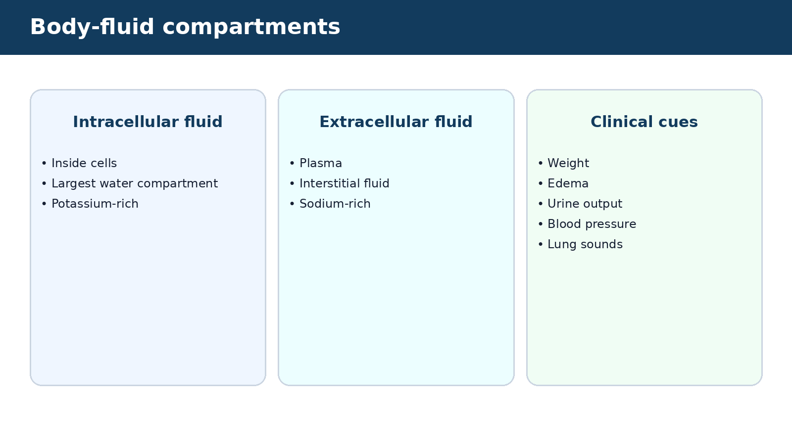
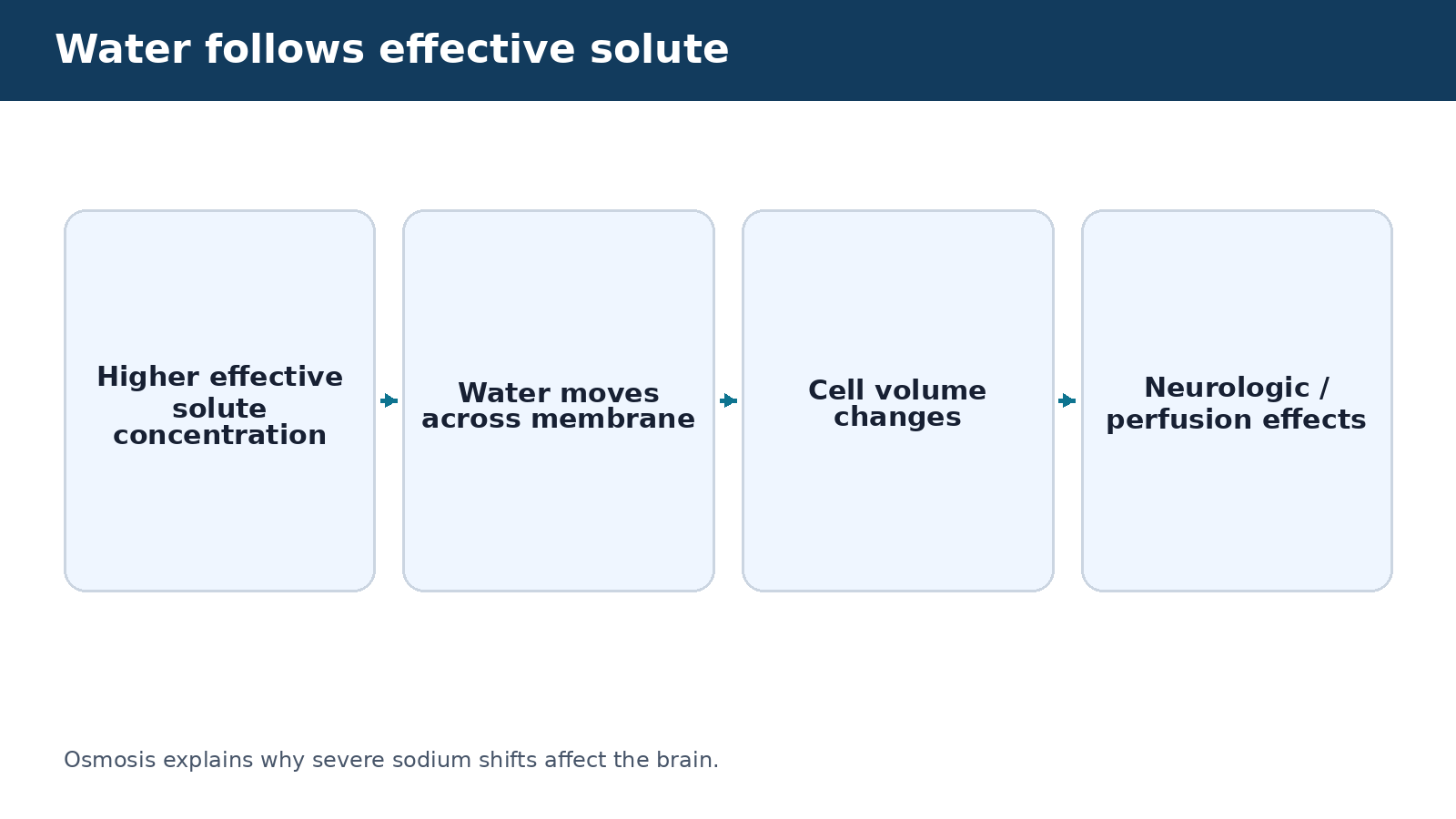
Direct answer: Body water is distributed mainly inside cells and in extracellular spaces. Water moves toward areas with a higher concentration of effective solute, while sodium is the major extracellular cation and potassium is the major intracellular cation.
A nursing student does not need a biochemistry degree to understand fluid balance. The practical question is where water is moving, what is causing the movement and how that movement affects circulation, cells and organs. The intracellular compartment holds most body water. Extracellular fluid includes plasma and interstitial fluid. Changes in sodium concentration strongly influence water movement between these compartments.
-
Osmosis is movement of water across a semipermeable membrane toward the side with higher effective solute concentration.
-
Diffusion is movement of particles down a concentration gradient.
-
Filtration moves fluid because of pressure differences, such as hydrostatic pressure across capillaries.
-
Albumin and other plasma proteins help create oncotic pressure that keeps water in the vascular space.
-
Kidneys, thirst, antidiuretic hormone and the renin-angiotensin-aldosterone system help regulate water and sodium balance.
Practical check: The bedside nurse sees these processes as edema, dry mucous membranes, weight change, blood-pressure change, urine-output change, lung sounds, mental-status change and laboratory trends.
Avoid this mistake: Do not equate edema with “too much total body fluid” in every case. A patient can have interstitial edema while effective circulating volume is low, such as in severe hypoalbuminemia or some forms of heart/liver failure.
NCLEX-RN® connection: NCLEX-RN® questions often give a fluid cue, an electrolyte value and a symptom. Connect the physiology to the immediate safety problem rather than memorizing isolated lists.
5. Assessing fluid status: combine history, examination, weight and trends
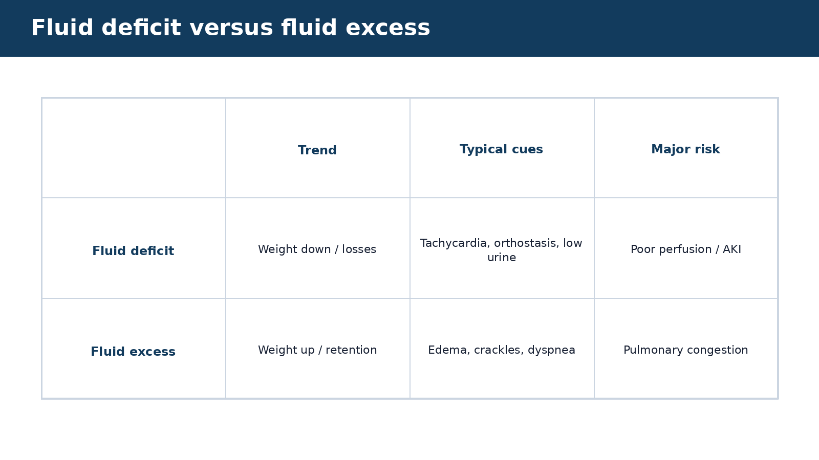
Direct answer: The most useful fluid assessment combines recent weight change, intake/output, urine output, blood pressure and pulse, edema, lung findings, mucous membranes, mental status and relevant laboratory trends.
-
No single sign perfectly proves fluid deficit or excess. Skin turgor, for example, can be less reliable in older adults. A nurse should compare multiple cues and the patient’s baseline.
-
Daily weight measured under similar conditions is often one of the most sensitive ways to follow fluid gain or loss. A change of about 1 kg represents roughly 1 liter of water, although body composition and measurement conditions matter.
-
Track intake from oral fluids, tube feedings, IV fluids, flushes, blood products and liquid medications when the clinical situation requires strict balance.
-
Track urine, drains, emesis, liquid stool and other measurable output. Insensible loss from skin and lungs is real but not usually measured directly.
-
New crackles, rising oxygen requirement, jugular venous distention, edema and rapid weight gain can suggest fluid excess in the right context.
-
Thirst, orthostasis, tachycardia, reduced urine output, dry mucosa and rising concentration of some laboratory values can support fluid deficit, but interpret them together.
Practical check: If urine output falls or weight changes quickly, ask whether the patient has also changed intake, diuretic therapy, renal function, hemodynamics or fluid losses.
Avoid this mistake: Do not diagnose dehydration from one dry mouth or overload from one ankle-swelling observation.
NCLEX-RN® connection: Reduction of Risk Potential includes recognizing trends and changes in condition. The trend is often more important than a single “normal” or “abnormal” number.
6. Tonicity and common IV fluids: what happens to water after infusion
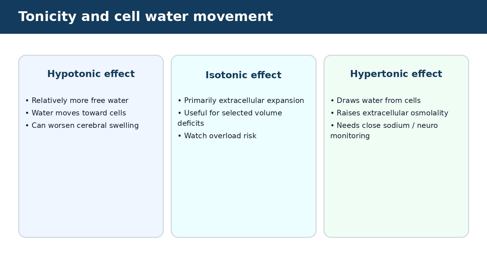
Direct answer: Isotonic solutions primarily expand extracellular volume, hypotonic solutions provide relatively more free water and shift water toward cells, and hypertonic solutions draw water out of cells into extracellular space. The patient’s diagnosis and sodium status determine whether that movement is helpful or dangerous.
Tonicity describes the effect of a solution on cell water movement. This is different from simply reading osmolarity printed on a bag. For example, D5W is isotonic in the bag, but once glucose is metabolized it behaves physiologically like free water.
Practical check: Fluid choice is a prescribing decision. The nurse verifies the solution, rate, indication and monitoring plan, then watches for respiratory distress, neurologic change, sodium shifts and access complications.
Avoid this mistake: Do not call D5W simply “isotonic” without explaining its physiologic behavior after glucose is metabolized.
NCLEX-RN® connection: Questions may test whether a fluid choice matches the clinical problem and whether a patient at risk for overload or cerebral swelling requires closer monitoring.
| Solution concept | Examples | Main teaching point |
|---|---|---|
| Isotonic | 0.9% sodium chloride; lactated Ringer solution | Expands extracellular/intravascular volume; monitor patients at risk of overload. |
| Hypotonic | 0.45% sodium chloride in selected situations | Provides proportionally more free water; can worsen cerebral edema or severe hyponatremia if used inappropriately. |
| Hypertonic | 3% sodium chloride for specific severe symptomatic hyponatremia protocols | Draws water from cells; requires close neurologic and sodium monitoring and careful correction. |
| Dextrose water nuance | D5W | Isotonic in container but becomes effectively hypotonic after glucose metabolism; not a resuscitation fluid for unstable volume loss. |
7. Sodium: understand water balance before memorizing symptoms
Direct answer: Serum sodium mainly reflects the relationship between total body sodium and water. Hyponatremia usually means there is too much water relative to sodium; hypernatremia usually means there is too little water relative to sodium. The brain is especially sensitive to rapid sodium changes.
-
A common adult laboratory reference interval is approximately 135–145 mEq/L (mmol/L), but use the reporting laboratory’s range. Sodium disorders are often disorders of water balance rather than simple dietary sodium deficiency or excess.
-
Hyponatremia may occur with excess water, diuretics, gastrointestinal losses replaced with free water, SIADH, adrenal problems, heart/liver/kidney disease and other causes.
-
Neurologic manifestations can include headache, confusion, lethargy, seizures and coma, especially when sodium falls rapidly or becomes severely low.
-
Hypernatremia often reflects water deficit from limited access to water, impaired thirst, fever, diarrhea, diabetes insipidus, osmotic diuresis or other losses.
-
Hypernatremia can cause thirst, irritability, weakness, confusion and seizures when severe or rapid.
-
Volume status matters: a patient with hyponatremia can be hypovolemic, euvolemic or hypervolemic, and treatment differs.
Practical check: Assess neurologic status, fluid balance, medications, glucose and the rate/direction of sodium change. Severe symptomatic sodium disorders require urgent management and frequent monitoring.
Avoid this mistake: Do not assume “low sodium = give salt” or “high sodium = restrict salt.” The cause and volume status determine treatment.
NCLEX-RN® connection: A new seizure or acute confusion with a markedly abnormal sodium level is a priority cue. Nursing questions focus on safety, monitoring and correct interpretation rather than prescribing a universal correction plan.
Sodium disorders: think water + brain
- Check sodium and its trend
- Assess neurologic symptoms and escalate urgent changes
- Determine volume status and possible causes
- Monitor the prescribed correction and repeat sodium results
8. Why sodium must often be corrected carefully
Direct answer: Rapid correction of chronic hyponatremia can cause osmotic demyelination, while overly rapid reduction of chronic hypernatremia can contribute to cerebral edema. Correction targets and frequency of monitoring depend on the cause, chronicity and symptoms and must follow current orders/guidelines.
Brain cells adapt to persistent changes in extracellular osmolality. That adaptation is protective over time but makes abrupt reversal dangerous. This is why severe sodium disorders are often managed with frequent laboratory checks and controlled therapy rather than a “normalize immediately” approach.
-
Acute severe symptomatic hyponatremia can be a neurologic emergency and may require hypertonic saline under a protocol with close monitoring.
-
Chronic or minimally symptomatic hyponatremia generally requires more cautious correction.
-
Hypernatremia from water deficit is usually corrected with water replacement at a rate chosen for the patient’s stability and chronicity.
-
Glucose can lower measured sodium by shifting water extracellularly; marked hyperglycemia changes interpretation and may require a corrected-sodium concept.
-
The nurse should watch for unexpected neurologic deterioration during treatment and promptly report a sodium trajectory that exceeds ordered goals.
Practical check: The nurse’s role is to verify fluid and rate, obtain scheduled labs, assess neurologic status and fluid balance, and escalate when the actual sodium trend differs from the planned trajectory.
Avoid this mistake: Do not memorize one universal “safe rate” and apply it to every patient. Guidelines use context-dependent limits, and high-risk patients may need even more conservative correction.
NCLEX-RN® connection: A student should recognize the danger of rapid correction even if exact prescriber-level targets are not being tested.
9. Potassium: the electrolyte with major electrical consequences
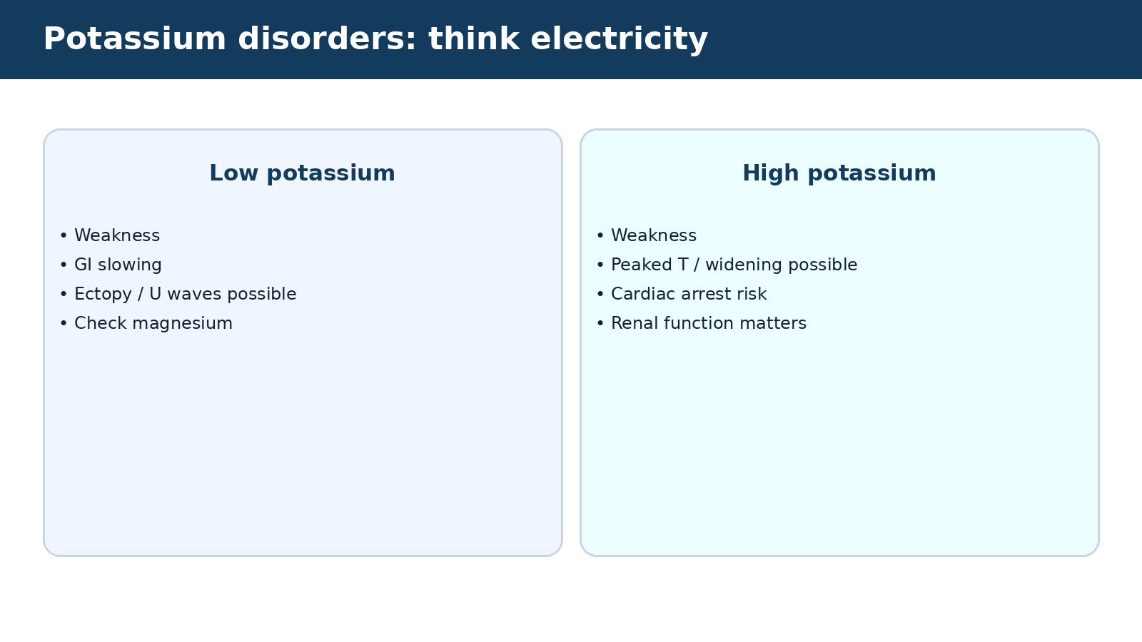
Direct answer: Potassium is essential for cardiac and skeletal-muscle electrical activity. A common adult reference interval is roughly 3.5–5.1 mEq/L, but the laboratory range should be used. Both low and high potassium can cause dangerous dysrhythmias.
-
Most potassium is inside cells, so small extracellular changes can have large electrical effects. Kidney function, acid-base status, insulin, tissue breakdown and medications all influence serum potassium.
-
Hypokalemia can result from gastrointestinal losses, diuretics, low intake, intracellular shifting from insulin or beta-agonists and other causes.
-
Findings may include weakness, cramps, constipation/ileus and ECG changes; severe hypokalemia can cause dysrhythmias.
-
Hyperkalemia can result from renal failure, potassium-sparing drugs, ACE inhibitor/ARB therapy, cell breakdown, acidosis, adrenal problems or excessive supplementation.
-
Hyperkalemia can cause weakness and ECG changes and may progress to lethal arrhythmia without dramatic early symptoms.
-
A falsely high potassium can result from hemolysis or specimen problems, so unexpected results should be correlated with the sample, ECG and clinical situation.
Practical check: For significant potassium abnormalities, review renal function, medications, recent replacements, acid-base status and ECG/telemetry when indicated.
Avoid this mistake: Do not assume a hemolyzed specimen can simply be ignored if the patient has risk factors or ECG changes; promptly obtain a reliable result while managing the clinical risk.
NCLEX-RN® connection: The combination of potassium abnormality plus ECG or muscle symptoms usually raises priority. The nurse should identify which finding requires urgent escalation.
10. Hypokalemia: replacement is important, but replacement itself can be dangerous
Direct answer: Potassium replacement may be oral or IV depending on severity, symptoms and ability to take enteral therapy. Potassium should never be given by direct IV push because a rapid concentrated dose can cause fatal dysrhythmia.
-
The safest nurse thinks about both deficiency and replacement risk. Before administering potassium, confirm the current value, order, renal function, urine output when relevant, route, concentration and infusion rate.
-
Oral replacement is often preferred when the patient is stable and the gastrointestinal route is usable.
-
IV potassium must be diluted and infused at a controlled rate according to order and institutional policy; higher-risk rates/concentrations require appropriate monitoring and access.
-
Inspect the IV site because potassium can irritate veins and extravasation can injure tissue.
-
Check magnesium when hypokalemia is persistent or difficult to correct because magnesium deficiency can promote renal potassium loss.
-
Identify ongoing losses such as diarrhea, vomiting, gastric suction or diuretic therapy.
Practical check: If a patient with hypokalemia has new weakness, palpitations or ECG changes, escalate promptly rather than focusing only on replacing a laboratory number.
Avoid this mistake: Never add potassium to a hanging IV bag at the bedside without pharmacy/policy safeguards, and never give IV push potassium.
NCLEX-RN® connection: “Never IV push potassium” is a high-yield safety rule. Questions may also test renal function, ECG monitoring and correction of coexisting magnesium deficiency.
11. Hyperkalemia: separate cardiac stabilization, shifting and potassium removal
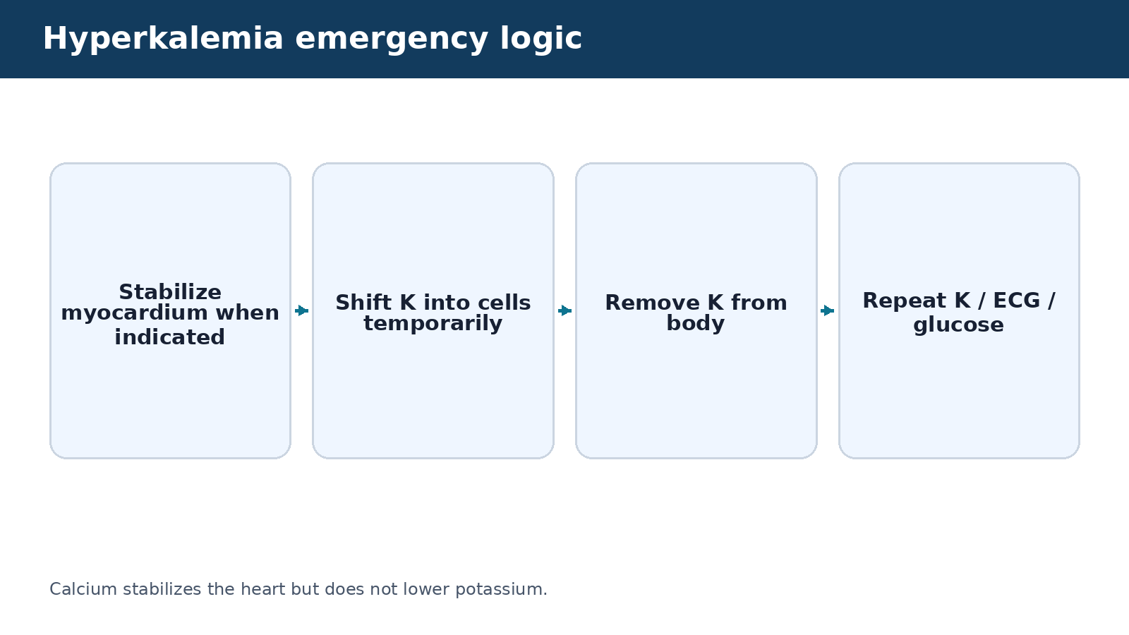
Direct answer: Emergency hyperkalemia management has three different goals: stabilize the myocardium when ECG toxicity is present, shift potassium into cells temporarily, and remove potassium from the body. These are not the same intervention.
Understanding the purpose of each intervention prevents a common reasoning error. IV calcium can protect the cardiac membrane but does not lower serum potassium. Insulin with glucose and some beta-agonists shift potassium into cells temporarily. Diuresis, gastrointestinal binders in selected contexts, or dialysis remove potassium from the body.
-
Place symptomatic or significant hyperkalemia on appropriate cardiac monitoring and obtain/interpret an ECG promptly.
-
Verify that glucose is monitored when insulin is used because hypoglycemia can occur.
-
Review kidney function and stop further potassium exposure when ordered/appropriate.
-
Recognize that improvement in ECG appearance after calcium does not mean the potassium concentration has been corrected.
-
Repeat potassium testing and reassessment according to the treatment plan because intracellular shifting is temporary.
Practical check: In a patient with severe hyperkalemia and ECG changes, this is a time-sensitive safety problem. Activate the appropriate urgent response rather than waiting for routine rounds.
Avoid this mistake: Do not say “calcium lowers potassium.” It stabilizes cardiac excitability. Do not assume one normal follow-up ECG means the hyperkalemia is resolved.
NCLEX-RN® connection: Items may ask which intervention protects the heart versus which lowers serum potassium. Knowing the purpose is more reliable than memorizing a medication list.
12. Calcium: total calcium, ionized calcium and neuromuscular function
Direct answer: Calcium supports bone structure, muscle contraction, nerve transmission and cardiac function. Total serum calcium is affected by albumin, while ionized calcium is the biologically active fraction.
-
A common total-calcium reference interval is around 8.5–10.5 mg/dL, but ranges vary. A low total calcium may partly reflect low albumin rather than true low ionized calcium. Clinical context and the laboratory’s reference range matter.
-
Hypocalcemia can occur with hypoparathyroidism, vitamin D deficiency, pancreatitis, kidney disease, massive transfusion from citrate binding and severe magnesium deficiency.
-
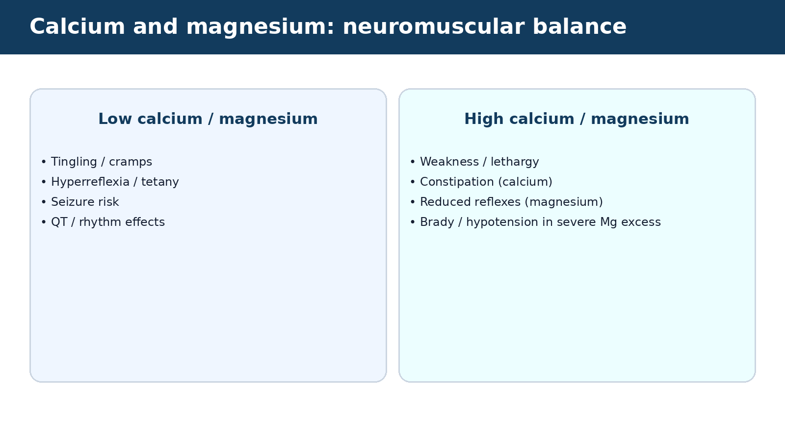
Symptoms can include perioral tingling, muscle cramps, tetany, seizures and prolonged QT-related dysrhythmia risk.
-
Hypercalcemia can occur with hyperparathyroidism, malignancy, medications and other causes.
-
Symptoms may include weakness, constipation, polyuria, dehydration, confusion and shortened QT in some cases.
-
Chvostek and Trousseau signs are classic teaching signs of neuromuscular irritability but should not replace broader clinical assessment.
Practical check: Assess symptoms, ECG when clinically indicated, renal function, phosphate/magnesium and the treatment context. Verify whether a total or ionized calcium result is being interpreted.
Avoid this mistake: Do not apply a “corrected calcium” formula blindly without understanding local laboratory practice and patient context; ionized calcium may be more useful in critically ill patients.
NCLEX-RN® connection: Hypocalcemia plus tetany/seizure is urgent. Hypercalcemia plus dehydration/mental-status change also requires focused assessment and treatment monitoring.
13. Magnesium: a quiet electrolyte with major cardiac and neuromuscular effects
Direct answer: Magnesium affects neuromuscular excitability, cardiac conduction and potassium/calcium regulation. Low magnesium can contribute to refractory hypokalemia and dysrhythmias; severe high magnesium can depress reflexes, blood pressure and respiration.
-
Serum magnesium reference ranges vary by laboratory. Many laboratories use values around 1.7–2.2 mg/dL, but always use the reported range and units because units can differ.
-
Hypomagnesemia can occur with diarrhea, malnutrition, alcohol use disorder, some diuretics, proton-pump inhibitors, renal losses and other causes.
-
Findings may include tremor, hyperreflexia, muscle cramps, seizures and dysrhythmias, including risk for torsades in the right context.
-
Hypermagnesemia is uncommon with normal kidney function and more often occurs when kidney clearance is impaired or magnesium-containing products accumulate.
-
High magnesium can cause nausea, lethargy, diminished deep-tendon reflexes, hypotension, bradycardia and respiratory depression as severity increases.
-
Magnesium replacement and magnesium-containing infusions require monitoring appropriate to the indication and dose.
Practical check: When potassium is not correcting as expected, check whether magnesium deficiency is contributing. When magnesium is high, review renal function and magnesium-containing medications/antacids/laxatives.
Avoid this mistake: Do not assume “more magnesium is harmless because it is a supplement.” Severe accumulation can be dangerous, especially with renal failure.
NCLEX-RN® connection: The nurse should connect magnesium abnormalities to cardiac rhythm, reflexes, breathing and coexisting electrolyte problems.
14. How sodium, potassium, calcium and magnesium interact
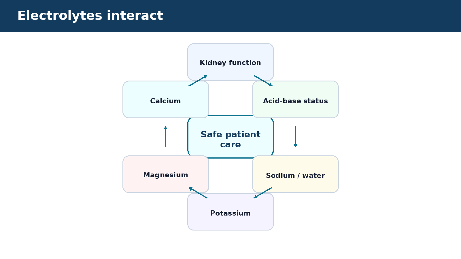
Direct answer: Electrolytes should not be interpreted as four unrelated numbers. Kidney function, acid-base status, hormones and one electrolyte abnormality can influence another.
The value of an electrolyte panel is pattern recognition. A patient with prolonged vomiting may have volume depletion, chloride loss, metabolic alkalosis and hypokalemia. A patient with diarrhea may have volume loss, potassium loss and metabolic acidosis. Kidney failure can produce fluid overload, hyperkalemia, acidosis and changes in calcium/phosphate balance.
-
Low magnesium can make potassium replacement ineffective because the kidneys continue losing potassium.
-
Calcium and magnesium both affect neuromuscular excitability; severe abnormalities may produce overlapping signs such as weakness, cramping or arrhythmia.
-
Acid-base shifts can change potassium distribution between intracellular and extracellular spaces.
-
Sodium concentration reflects water balance, while total-body sodium determines extracellular volume; these are related but not identical concepts.
-
Kidney dysfunction can change multiple electrolytes at once, so trends in creatinine and urine output matter.
Practical check: When one electrolyte is abnormal, scan the rest of the metabolic panel and the clinical story rather than treating the value in isolation.
Avoid this mistake: Avoid “one lab = one symptom” thinking. A confused patient with low sodium may also be septic, hypoglycemic or taking sedating medications.
NCLEX-RN® connection: Clinical judgment questions reward cue clustering: connect electrolytes with ECG, neurologic findings, medications, intake/output and kidney function.
15. Chloride, bicarbonate and phosphate: supporting electrolytes worth understanding
Direct answer: Chloride and bicarbonate help explain acid-base and volume patterns, while phosphate is important for ATP, muscle function and bone. These are not the headline electrolytes in this guide, but they help make the pattern coherent.
-
A basic metabolic panel usually includes sodium, potassium, chloride and bicarbonate/total CO2 along with glucose and kidney markers. Chloride often moves with sodium but can change independently in acid-base disorders.
-
Low chloride commonly accompanies vomiting or gastric suction and can support metabolic alkalosis.
-
High chloride may accompany large chloride-rich fluid loads or non-anion-gap metabolic acidosis.
-
Low bicarbonate suggests metabolic acidosis or respiratory alkalosis compensation; high bicarbonate suggests metabolic alkalosis or respiratory acidosis compensation, but ABG context may be needed.
-
Severe hypophosphatemia can impair respiratory-muscle and cardiac function, while hyperphosphatemia is common in kidney failure.
-
Laboratory reference ranges vary, and serum levels may not reflect total-body stores perfectly.
Practical check: Use these values to support a pattern, then assess the patient and underlying cause.
Avoid this mistake: Do not diagnose a full acid-base disorder from serum bicarbonate alone when the clinical situation requires blood-gas interpretation.
NCLEX-RN® connection: Questions may combine vomiting, diarrhea, NG suction, kidney failure or refeeding risk with electrolyte data.
16. Fluid deficit: recognize reduced circulating volume and its causes
Direct answer: Fluid-volume deficit means extracellular volume has fallen enough to affect perfusion or hydration. Causes include gastrointestinal loss, bleeding, diuresis, fever, poor intake and third spacing, among others.
-
The exact label “dehydration” is often used loosely. Clinically, distinguish water deficit from loss of isotonic extracellular fluid and from blood loss because treatment differs.
-
Possible cues: tachycardia, orthostatic symptoms, low blood pressure, reduced urine output, dry mucosa, concentrated urine, delayed capillary refill, weakness and acute kidney injury.
-
Bleeding adds loss of red-cell mass and can be occult; do not assume all low-volume states are simple dehydration.
-
Older adults and patients with cognitive/functional limitations may not sense or obtain water effectively.
-
Diuretics, osmotic diuresis and high-output drains can produce rapid losses.
-
Treatment requires addressing the cause and using the prescribed fluid/blood strategy; fluid choice depends on the problem.
Practical check: Trend mental status, urine output, hemodynamics, weight and response to replacement. Watch high-risk patients for fluid overload during correction.
Avoid this mistake: Do not give large amounts of fluid automatically to every hypotensive patient; cardiogenic shock, obstructive shock and severe heart failure require different decisions.
NCLEX-RN® connection: Ask why the patient is volume depleted and whether the current signs suggest compromised perfusion.
17. Fluid excess: recognize congestion before it becomes respiratory failure
Direct answer: Fluid-volume excess can produce edema and pulmonary congestion when intake or sodium/water retention exceeds the body’s ability to excrete or distribute fluid safely.
-
Common contexts include heart failure, kidney failure, excessive IV fluid, liver disease and some endocrine states. Peripheral edema alone does not define severity; pulmonary symptoms and oxygenation often determine urgency.
-
High-yield cues include rapid weight gain, increasing edema, crackles, dyspnea, rising oxygen requirement, jugular venous distention and hypertension in some patients.
-
Fluid restriction and sodium management may be part of the plan for selected patients, but exact limits are patient specific.
-
Diuretics require monitoring of urine output, blood pressure, kidney function and electrolytes.
-
A patient with new pulmonary edema may require urgent oxygen/ventilatory support and treatment of the underlying cause.
-
Daily weight and trend-based assessment can detect worsening congestion earlier than waiting for severe shortness of breath.
Practical check: If a patient on IV fluids develops new dyspnea, crackles or oxygen need, reassess the infusion and escalate promptly rather than simply charting “fluid overload.”
Avoid this mistake: Do not use ankle edema alone to decide whether to stop prescribed fluids; assess the whole patient and follow orders/escalation.
NCLEX-RN® connection: Breathing deterioration takes priority over routine intake/output completion.
18. Diuretics and electrolyte monitoring
Direct answer: Diuretics can improve congestion but may cause volume depletion, kidney injury and electrolyte changes. The pattern depends on the drug class and patient.
-
Loop and thiazide-type diuretics commonly lower potassium and sometimes magnesium, while potassium-sparing agents can increase potassium. All can contribute to hypotension or kidney-function changes in the wrong context.
-
Review blood pressure, symptoms, urine output, weight and relevant electrolytes/renal function.
-
Teach patients to report severe weakness, palpitations, dizziness or signs of dehydration.
-
Schedule and dietary instructions depend on the specific medication and plan; do not generalize one teaching rule to all diuretics.
-
Monitor for orthostasis and fall risk, especially in older adults or after dose changes.
-
When diuresis is aggressive, trend potassium and magnesium because dysrhythmia risk can increase.
Practical check: The goal is not simply “more urine.” It is improved congestion without creating dangerous volume or electrolyte depletion.
Avoid this mistake: Do not assume every diuretic causes hypokalemia. Potassium-sparing diuretics can cause hyperkalemia.
NCLEX-RN® connection: Medication questions often pair a diuretic with potassium, blood pressure or renal data and ask whether the medication can be given safely.
19. Electrolytes and the ECG: when a lab value becomes an electrical emergency
Direct answer: Potassium has the strongest direct connection to life-threatening ECG changes, but calcium and magnesium can also affect intervals and rhythm. Treat the patient and ECG, not only the printed laboratory value.
-
ECG manifestations are not perfectly predictable and can be absent despite dangerous electrolyte levels. A normal-looking ECG does not prove safety, especially with significant hyperkalemia.
-
Hyperkalemia can produce peaked T waves, PR prolongation, QRS widening and progressive conduction abnormalities as severity increases.
-
Hypokalemia can cause ST depression, flattened T waves, prominent U waves and ventricular ectopy.
-
Hypocalcemia tends to prolong the QT interval; hypercalcemia tends to shorten it.
-
Low magnesium increases risk for ventricular dysrhythmias and torsades in susceptible patients.
-
Telemetry monitoring should be used when clinically indicated by severity, symptoms or treatment plan.
Practical check: If a patient with an electrolyte abnormality develops palpitations, syncope, chest symptoms or new rhythm changes, escalate urgently.
Avoid this mistake: Do not wait for the “classic” ECG pattern before treating a clinically dangerous electrolyte disorder.
NCLEX-RN® connection: Questions may ask which laboratory result is most urgent in a patient with ECG changes. Potassium and symptomatic calcium/magnesium abnormalities should trigger safety-focused reasoning.
20. Specimen quality and pseudoelectrolyte abnormalities
Direct answer: Before acting on an unexpected laboratory value, consider whether the specimen is reliable—but never delay urgent care when the patient’s clinical picture supports the abnormality.
-
Hemolysis can release intracellular potassium and create pseudohyperkalemia. Prolonged tourniquet use, sample contamination, drawing near an infusion and collection/processing delays can also alter results.
-
Compare the new result with previous values and the patient’s symptoms.
-
Check whether the laboratory flagged hemolysis or sample quality.
-
If a result is unexpected and the patient is stable, promptly repeat it using correct collection technique as ordered/policy.
-
If the patient has ECG changes or severe symptoms, treat the situation as potentially real while confirmation is obtained.
-
Do not draw from an IV line infusing electrolytes or fluids unless the approved technique prevents contamination and the order/policy allows it.
Practical check: Good nursing judgment balances two risks: treating a false result and ignoring a true emergency.
Avoid this mistake: Do not dismiss a high potassium merely because hemolysis is possible.
NCLEX-RN® connection: A question may include both a hemolyzed specimen note and an asymptomatic patient; the best action often involves repeat testing rather than immediate assumption.
21. A practical electrolyte interpretation sequence
Direct answer: Use the same sequence every time: verify the value and units, compare with trend, assess symptoms, check kidney function and medications, look for ECG/neurologic risk, identify the likely cause, then evaluate the treatment response.
This sequence turns a lab panel into clinical reasoning. It prevents the common mistake of jumping directly from “low” or “high” to a memorized intervention.
-
Verify: correct patient, sample, units and reference range.
-
Trend: acute change or long-standing pattern?
-
Symptoms: neurologic, cardiac, respiratory, gastrointestinal or muscular changes?
-
Context: intake/output, renal function, GI loss, medications, endocrine disease, acid-base status.
-
Immediate risk: dysrhythmia, seizure, severe weakness, respiratory depression or unstable hemodynamics?
-
Plan: ordered replacement/restriction/removal strategy, monitoring frequency and route.
-
Reassess: repeat laboratory result plus patient response.
Practical check: This approach works for sodium, potassium, calcium and magnesium and supports the six NCJMM clinical-judgment steps.
Avoid this mistake: Do not “treat the lab” without deciding whether the patient is stable and whether the value is real.
NCLEX-RN® connection: When options are similar, the safest answer usually collects or acts on the cue that changes immediate risk.
Electrolyte interpretation sequence
- Verify patient, specimen, value, units and reference range
- Compare trends and assess symptoms
- Review kidney function, medications and fluid balance
- Escalate dangerous ECG, neurologic or hemodynamic findings immediately
- Follow the prescribed treatment and reassess laboratory values and symptoms
22. Clinical judgment cases
How to use the cases: Read the scenario once for the big picture. Then identify the most important cues, the priority concern, the safest immediate nursing actions, and the findings that would show whether the plan worked. These cases are original educational examples, not official examination items.
Case 1: Hyponatremia with new confusion
An older adult receiving a thiazide-type diuretic has sodium 122 mEq/L, new confusion and headache. The previous sodium was 136 mEq/L.
| Clinical-judgment step | Reasoning |
|---|---|
| Recognize cues | Marked sodium decline plus acute neurologic symptoms. |
| Analyze cues | The patient may have symptomatic hyponatremia with a meaningful acute change; medication and fluid balance are relevant causes. |
| Prioritize hypotheses | Neurologic safety and urgent evaluation take priority. |
| Generate solutions | Institute safety precautions, perform focused neurologic assessment, notify urgently, review medications/intake and prepare for ordered controlled correction with frequent sodium monitoring. |
| Take action | Escalate the symptomatic sodium change rather than encouraging free water or waiting for routine review. |
| Evaluate outcomes | Trend neurologic status and serum sodium at the ordered frequency and watch for overly rapid correction. |
Case 2: Hyperkalemia and a widening QRS
A patient with kidney failure has potassium 6.8 mEq/L and new QRS widening on the ECG.
| Clinical-judgment step | Reasoning |
|---|---|
| Recognize cues | Severe hyperkalemia with ECG toxicity. |
| Analyze cues | The patient is at immediate risk of malignant dysrhythmia. |
| Prioritize hypotheses | Cardiac stabilization and urgent hyperkalemia treatment. |
| Generate solutions | Activate urgent care, place on continuous monitoring, obtain treatment orders/protocol interventions that stabilize myocardium, shift potassium and remove it, and monitor glucose when insulin is used. |
| Take action | Do not wait for routine repeat labs before responding to the ECG change. |
| Evaluate outcomes | Repeat ECG, potassium and glucose and monitor for recurrent hyperkalemia after temporary shifting therapies. |
Case 3: Hypokalemia after gastrointestinal losses
A patient has repeated vomiting and NG suction. Potassium is 2.9 mEq/L; the patient reports weakness and palpitations.
| Clinical-judgment step | Reasoning |
|---|---|
| Recognize cues | Ongoing GI loss, low potassium and cardiac symptoms. |
| Analyze cues | The deficit may continue unless losses are addressed; magnesium may also be low. |
| Prioritize hypotheses | Assess rhythm and replace potassium safely while identifying cause. |
| Generate solutions | Obtain ECG/monitoring as indicated, review magnesium and renal function, verify ordered replacement and treat ongoing losses. |
| Take action | Administer ordered replacement by a safe route/rate; never IV push potassium. |
| Evaluate outcomes | Recheck symptoms, ECG and potassium/magnesium after replacement. |
Case 4: Fluid overload during IV therapy
A patient with chronic heart failure receiving IV fluids develops new dyspnea, bilateral crackles and increasing oxygen requirement.
| Clinical-judgment step | Reasoning |
|---|---|
| Recognize cues | Respiratory deterioration while receiving fluid. |
| Analyze cues | Pulmonary congestion/fluid overload is possible; other causes of dyspnea must also be considered. |
| Prioritize hypotheses | Support breathing and prevent further worsening. |
| Generate solutions | Sit patient upright if appropriate, assess oxygenation and hemodynamics, pause/reassess infusion per protocol and urgently notify for treatment. |
| Take action | Address respiratory status immediately rather than waiting for the next daily weight. |
| Evaluate outcomes | Track oxygenation, work of breathing, lung findings, urine output and weight after treatment. |
Case 5: Hypermagnesemia in renal failure
A patient with advanced kidney failure has been using magnesium-containing laxatives and now has lethargy, hypotension and diminished reflexes.
| Clinical-judgment step | Reasoning |
|---|---|
| Recognize cues | Renal failure plus magnesium exposure and classic neuromuscular/hemodynamic depression. |
| Analyze cues | Magnesium accumulation may be causing clinically significant hypermagnesemia. |
| Prioritize hypotheses | Airway/breathing/hemodynamic monitoring and urgent management. |
| Generate solutions | Stop further magnesium exposure, obtain urgent magnesium/renal labs and ECG, notify and prepare for ordered antagonism/removal measures. |
| Take action | Escalate immediately because respiratory depression can develop. |
| Evaluate outcomes | Monitor reflexes, respiratory status, blood pressure, ECG and magnesium level. |
Case 6: Calcium change after massive transfusion
A patient receiving a large-volume transfusion develops tingling, muscle twitching and a prolonged QT interval.
| Clinical-judgment step | Reasoning |
|---|---|
| Recognize cues | Neuromuscular irritability and QT prolongation in a context where citrate can lower ionized calcium. |
| Analyze cues | Acute hypocalcemia is possible. |
| Prioritize hypotheses | Prevent dysrhythmia/seizure and maintain perfusion. |
| Generate solutions | Obtain/verify ionized calcium as ordered, monitor ECG, notify urgently and prepare for prescribed calcium replacement. |
| Take action | Do not dismiss symptoms as anxiety in this high-risk context. |
| Evaluate outcomes | Reassess symptoms, ECG and ionized calcium after treatment. |
Go deeper: Apply the same approach to Next Generation NCLEX® case studies.
23. Practice questions with answers and rationales
Important: These are original RN Clarity practice questions. They are not copied from, endorsed by or affiliated with NCSBN® or the NCLEX® examination.
Question 1: Which finding is most useful for following day-to-day fluid gain or loss when measured consistently?
-
A. Daily body weight
-
B. One skin-turgor check
-
C. One serum sodium value
-
D. Height
Answer and rationale: A. Consistent daily weight is highly sensitive to fluid change. It should still be interpreted with intake/output and clinical findings.
Question 2: Which statement best describes serum sodium?
-
A. It always measures total body sodium stores directly.
-
B. It primarily reflects the relationship between body water and sodium.
-
C. Low sodium always means the patient needs more dietary salt.
-
D. High sodium always means the patient has fluid overload.
Answer and rationale: B. Serum sodium is a concentration and commonly reflects water balance relative to sodium. Volume status must be assessed separately.
Question 3: A patient with severe hyperkalemia has ECG changes. Which ordered intervention stabilizes the cardiac membrane but does not lower serum potassium?
-
A. IV calcium
-
B. Insulin with glucose
-
C. A potassium-removal therapy
-
D. A loop diuretic when appropriate
Answer and rationale: A. Calcium antagonizes the cardiac membrane effects of hyperkalemia but does not remove potassium or shift it into cells.
Question 4: Which action is unsafe for potassium replacement?
-
A. Verify renal function.
-
B. Use a controlled IV infusion when IV replacement is ordered.
-
C. Give concentrated potassium by direct IV push.
-
D. Recheck potassium after replacement.
Answer and rationale: C. IV push potassium can cause fatal dysrhythmia and should never be given.
Question 5: Why might hypokalemia be difficult to correct when magnesium is also low?
-
A. Magnesium deficiency can promote continued renal potassium loss.
-
B. Magnesium always raises potassium directly.
-
C. Low magnesium prevents potassium measurement.
-
D. They are unrelated.
Answer and rationale: A. Magnesium deficiency can make potassium repletion refractory by increasing renal potassium wasting.
Question 6: Which symptom is especially concerning in severe hyponatremia?
-
A. Mild dry skin
-
B. New seizure
-
C. Stable chronic ankle edema
-
D. Occasional hiccup
Answer and rationale: B. Severe or rapidly developing hyponatremia can cause cerebral edema and neurologic emergencies including seizure.
Question 7: A patient with heart failure on IV fluids develops crackles and increased oxygen need. What is the nurse’s priority?
-
A. Finish the ordered bag before notifying anyone.
-
B. Assess and support respiratory status and escalate possible fluid overload.
-
C. Encourage additional oral fluids.
-
D. Wait for the morning weight.
Answer and rationale: B. New breathing impairment is an acute priority and may indicate pulmonary congestion.
Question 8: Which statement about D5W is most accurate?
-
A. It remains strongly hypertonic after infusion.
-
B. It is isotonic in the bag but provides free water after glucose is metabolized.
-
C. It is the preferred shock-resuscitation fluid in every patient.
-
D. It contains no water.
Answer and rationale: B. D5W becomes effectively hypotonic after glucose metabolism and should not be treated as a universal volume-resuscitation fluid.
Question 9: Which patient cue best supports hypermagnesemia?
-
A. Hyperreflexia and tremor
-
B. Diminished reflexes with hypotension and lethargy
-
C. Severe thirst only
-
D. Peaked T waves as the only possible sign
Answer and rationale: B. Significant magnesium excess can depress neuromuscular function and cause hypotension, bradycardia and respiratory depression.
Question 10: Which ECG change is commonly associated with hypocalcemia?
-
A. Shortened QT
-
B. Prolonged QT
-
C. Always a narrow QRS with peaked T waves
-
D. No possible ECG effect
Answer and rationale: B. Hypocalcemia can prolong the QT interval, while hypercalcemia tends to shorten it.
Question 11: An unexpectedly high potassium result is flagged as hemolyzed and the patient is stable without ECG changes. What is a reasonable next step?
-
A. Assume the patient has lethal hyperkalemia without confirmation.
-
B. Ignore the result permanently.
-
C. Promptly obtain a properly collected repeat specimen while considering the clinical context.
-
D. Give potassium replacement.
Answer and rationale: C. Hemolysis can cause pseudohyperkalemia. A stable patient with discordant data needs timely confirmation, while clinical deterioration would require urgent action.
Question 12: Which patient is at greatest risk for hypermagnesemia?
-
A. A healthy adult with normal kidneys eating magnesium-rich food
-
B. A patient with advanced kidney failure taking magnesium-containing products
-
C. A patient with mild seasonal allergies
-
D. A runner who drinks water
Answer and rationale: B. Kidneys clear magnesium. Accumulation is more likely when renal function is severely impaired and magnesium exposure continues.
Question 13: What is the best interpretation of edema?
-
A. It always proves intravascular volume excess.
-
B. It shows fluid has accumulated in tissues, but effective circulating volume may still be low.
-
C. It always means sodium is high.
-
D. It rules out dehydration.
Answer and rationale: B. Interstitial edema can coexist with low effective circulating volume, so the nurse must assess the whole fluid-distribution pattern.
Question 14: Which action is appropriate when a patient with hypokalemia receives IV potassium?
-
A. Administer as a rapid IV push.
-
B. Use the ordered dilution/rate and monitor the IV site and patient.
-
C. Mix it into any hanging bag without a label.
-
D. Skip renal-function review.
Answer and rationale: B. IV potassium requires controlled dilution/rate and monitoring because rapid or concentrated administration can be dangerous.
Question 15: Which statement about hypertonic saline is correct?
-
A. It can be used casually for mild asymptomatic hyponatremia.
-
B. It may be used in selected severe symptomatic hyponatremia protocols with close monitoring.
-
C. It always lowers sodium.
-
D. It is identical to free water.
Answer and rationale: B. Hypertonic saline raises sodium and osmolality and is reserved for selected indications with careful correction monitoring.
Question 16: Which pattern best fits fluid-volume deficit?
-
A. Rapid weight gain, crackles and edema
-
B. Orthostatic symptoms, tachycardia and reduced urine output
-
C. JVD and increasing oxygen need
-
D. New ascites with no other data always
Answer and rationale: B. Orthostasis, tachycardia and low urine output are compatible with reduced effective circulating volume when interpreted together.
Question 17: Which statement about laboratory reference ranges is safest?
-
A. The same exact range applies in every laboratory.
-
B. Use the reporting laboratory’s reference range and units while understanding common expected values.
-
C. Reference ranges do not matter.
-
D. Only textbook ranges are valid.
Answer and rationale: B. Methods and units can differ. Nursing interpretation should use the actual lab’s range and the patient’s clinical context.
Question 18: Which symptom after severe hypernatremia treatment would require urgent reassessment?
-
A. New neurologic deterioration
-
B. Stable appetite
-
C. A normal pulse
-
D. Request for water
Answer and rationale: A. Neurologic change during correction can signal dangerous osmotic shifts and requires prompt evaluation.
Question 19: A patient taking a potassium-sparing diuretic should be monitored for which electrolyte problem?
-
A. Hyperkalemia
-
B. Hypokalemia only
-
C. Hypernatremia always
-
D. Hypocalcemia only
Answer and rationale: A. Potassium-sparing diuretics can increase serum potassium, especially with kidney dysfunction or interacting therapies.
Question 20: What is the best first question when an electrolyte value is abnormal?
-
A. Which memorized replacement should I give?
-
B. Is the value reliable, how has it changed, and what is happening clinically?
-
C. Can I ignore it if the patient feels fine?
-
D. Should all IV fluids be stopped?
Answer and rationale: B. Safe interpretation begins with reliability, trend and symptoms before selecting or anticipating treatment.
Go deeper: Continue with NCLEX-RN® practice questions.
24. Frequently asked questions
What are normal sodium and potassium levels?
Common adult reference ranges are about 135–145 mEq/L for sodium and 3.5–5.1 mEq/L for potassium, but laboratories use slightly different ranges. Always interpret the patient’s actual report and units.
What is the difference between dehydration and fluid-volume deficit?
“Dehydration” often refers to water deficit, while fluid-volume deficit commonly describes loss of extracellular/intravascular volume. In everyday clinical language the terms may overlap, but the cause and sodium/volume pattern matter for treatment.
Why can hyponatremia cause seizures?
A rapid or severe fall in extracellular osmolality can move water into brain cells and cause cerebral edema, producing headache, confusion, seizure or coma.
Why can sodium correction be dangerous?
The brain adapts to chronic osmolality changes. Correcting chronic hyponatremia too rapidly can cause osmotic demyelination; correcting chronic hypernatremia too rapidly can contribute to cerebral edema.
Why is potassium so important for nurses?
Potassium strongly affects cardiac conduction and muscle function. Both severe hypokalemia and hyperkalemia can produce life-threatening dysrhythmias.
Can IV calcium treat hyperkalemia?
IV calcium can stabilize the myocardium when hyperkalemia is causing ECG toxicity, but it does not lower serum potassium. Other therapies shift or remove potassium.
Why check magnesium with low potassium?
Magnesium deficiency can make hypokalemia difficult to correct because renal potassium losses continue.
What is the best indicator of fluid change?
No single sign is perfect, but consistently measured daily weight is very useful. Combine it with intake/output, urine output, hemodynamics, examination and lab trends.
Is D5W isotonic or hypotonic?
It is approximately isotonic in the IV bag, but after glucose is metabolized the remaining water behaves as free water, making its physiologic effect hypotonic.
Can edema occur when a patient has low circulating volume?
Yes. Fluid can be trapped in interstitial or third spaces while effective vascular volume is low, such as in severe hypoalbuminemia or some advanced heart/liver conditions.
Should nurses memorize exact electrolyte replacement doses?
Students should understand indications, safety principles and monitoring. Actual replacement doses, concentrations and rates are patient specific and must come from current orders, protocols and references.
How does this topic appear on NCLEX-RN® questions?
Often through cue clustering: an electrolyte result plus ECG, neurologic, renal, medication or fluid-balance findings. The safest answer identifies the immediate risk and appropriate nursing monitoring/escalation.
25. Official and primary references
Sources checked September 10, 2026. Use the linked guidance for the full context and follow patient-specific orders, scope of practice and local policy.
-
2026 NCLEX-RN® Test Plan — Official blueprint for fluid/electrolyte assessment, IV therapy, medications and risk reduction.
-
Electrolyte Panel - MedlinePlus — NIH/NLM overview of sodium, potassium, chloride and bicarbonate testing.
-
Potassium Blood Test - MedlinePlus — Potassium function and interpretation.
-
Low Blood Potassium - MedlinePlus — Common potassium reference range and clinical overview.
-
Calcium Blood Test - MedlinePlus — Calcium testing and common causes of abnormal results.
-
Magnesium Blood Test - MedlinePlus — Magnesium physiology and abnormal-level overview.
-
Basic Metabolic Panel - MedlinePlus — Fluid balance, electrolytes and kidney-function testing context.
26. Educational, clinical-safety and trademark disclaimer
Educational and clinical-safety disclaimer: This guide is provided by RN Clarity for general educational and informational purposes. It does not replace the official 2026 NCLEX-RN® Test Plan, nursing-school instruction, a current drug or clinical reference, provider orders, nursing regulator requirements, institutional policy, manufacturer instructions, local infection-control guidance or professional clinical judgment. Real patients may have conditions, medications, allergies, laboratory results, age-related needs or other factors that require a different approach. When a patient may be deteriorating, follow local emergency and escalation procedures. Use of this guide does not guarantee examination success or clinical competence. Non-affiliation and trademark notice: RN Clarity is an independent educational resource and is not affiliated with, endorsed by, sponsored by, approved by or officially connected with the National Council of State Boards of Nursing, Inc. NCLEX®, NCLEX-RN®, NCLEX-PN® and NCSBN® are registered trademarks of the National Council of State Boards of Nursing, Inc. All trademarks belong to their respective owner. Their use here is for identification, commentary and educational reference only.