ECG Interpretation for Nurses: 24 Rhythms & Nursing Actions
Learn ECG and EKG interpretation step by step. Understand rate, rhythm, intervals, 24 rhythm strips, causes, urgency and priority nursing actions — aligned to the 2026 NCLEX-RN® Test Plan.
Quick Answer: What Is the Fastest Way to Interpret an ECG Strip?
Nurses use an eight-step systematic method to interpret any ECG or telemetry strip: determine rate, assess regularity, examine P waves, measure the PR interval, measure the QRS width, examine the ST segment, measure the QT interval, and state the interpretation. Applying the same steps in the same order on every strip eliminates omission errors and produces a defensible nursing assessment. On the NCLEX-RN®, rhythm identification is tested as a clinical judgment skill — the correct answer always connects the ECG finding to the nursing action it requires.
Quick Answer: The first step for any new arrhythmia is to assess the client's hemodynamic status — pulse, blood pressure, level of consciousness, chest pain, and signs of perfusion. An ECG finding alone does not determine the nursing response; the client's tolerance determines urgency.
Go deeper: For how ECG findings function as clinical cues inside the CJMM six-step framework, see the clinical judgment guide.
1. Why ECG Matters for NCLEX-RN®
Electrocardiogram (ECG) interpretation is one of the highest-yield clinical skills tested on the NCLEX-RN® because cardiac dysrhythmias appear across multiple Client Needs categories. Under the April 2026 Test Plan, ECG content surfaces in Physiological Adaptation, Reduction of Risk Potential, and Pharmacological and Parenteral Therapies — together representing more than 30% of examination items.
Entry-level registered nurses working in medical-surgical, telemetry, critical care, emergency, postpartum, or pediatric settings are expected to:
- Recognize normal sinus rhythm and common dysrhythmias on a monitor or rhythm strip.
- Identify findings that require immediate action versus those that need provider notification within a defined window.
- Connect ECG changes to electrolyte imbalances, medications, and clinical conditions.
- Initiate appropriate nursing interventions based on the client's hemodynamic response.
- Document rhythm strips accurately and communicate findings using structured handoffs.
The NCLEX-RN® does not expect nurses to perform advanced electrophysiology interpretation. The examination focuses on recognition of 24 commonly tested rhythms, understanding of the eight-step method, correlation of ECG changes with common causes, and selection of correct nursing priorities.
Go deeper: For how the April 2026 NCLEX-RN® Test Plan organizes content areas and clinical judgment integration, see the NCLEX-RN® test plan explained.
2. Electrical Conduction of the Heart
Understanding the normal conduction pathway is essential before interpreting any dysrhythmia, because every abnormal rhythm represents a deviation from this pathway.
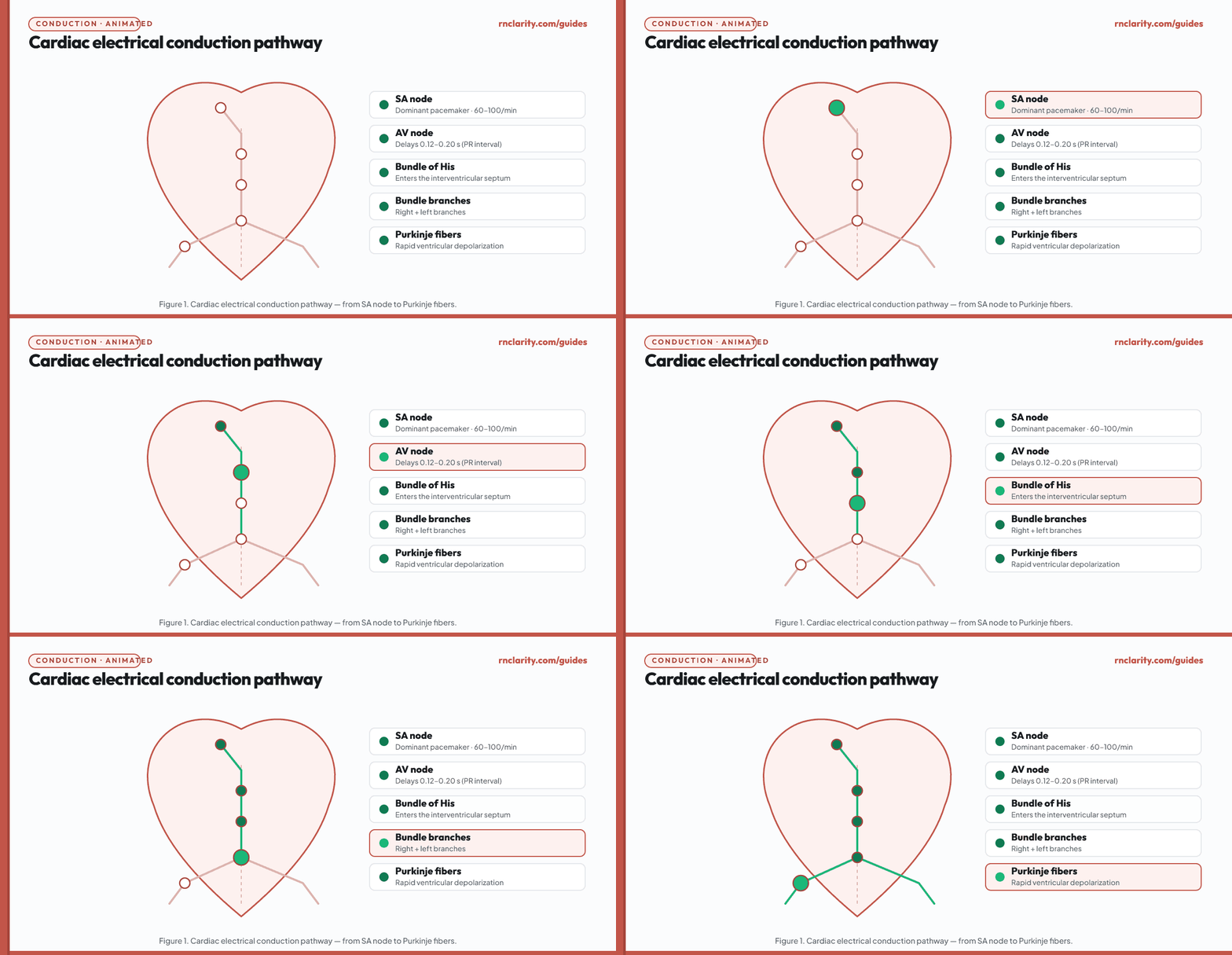
2.1 Normal conduction sequence
The sinoatrial (SA) node, located in the upper right atrium, is the dominant pacemaker of the heart. It generates impulses at an intrinsic rate of 60–100 beats per minute. From the SA node, the impulse spreads through the atrial myocardium via internodal pathways, depolarizing both atria simultaneously — this atrial depolarization produces the P wave on the ECG.
The impulse reaches the atrioventricular (AV) node at the junction of the right atrium and right ventricle. The AV node intentionally delays conduction for approximately 0.12–0.20 seconds, allowing the atria to complete contraction and fill the ventricles before ventricular systole begins. This delay is visible on the ECG as the PR interval.
From the AV node, the impulse enters the bundle of His, then divides into the right bundle branch and the left bundle branch. The left bundle branch divides further into the left anterior fascicle and left posterior fascicle. Bundle branches carry the impulse rapidly to the Purkinje fiber network, which distributes it simultaneously throughout the ventricular myocardium, causing rapid depolarization — the QRS complex on the ECG.
Ventricular repolarization (recovery) follows and is represented by the T wave. The ST segment between the QRS complex and the T wave normally lies at or near the isoelectric baseline.
2.2 Backup pacemaker hierarchy
If the SA node fails or its impulse is blocked, subsidiary pacemakers take over at slower intrinsic rates:
| Pacemaker site | Intrinsic rate | ECG consequence |
|---|---|---|
| SA node | 60–100/min | Normal sinus rhythm; upright P waves in lead II. |
| AV node / junction | 40–60/min | Junctional rhythm; retrograde or absent P waves. |
| Bundle branches / Purkinje fibers | 20–40/min | Idioventricular rhythm; wide, bizarre QRS; life-threatening rate. |
Understanding this hierarchy explains why third-degree (complete) AV block can produce a wide-complex ventricular escape rhythm — the ventricles are driven by the lowest, slowest backup pacemaker.
2.3 Mechanisms of dysrhythmia
Three fundamental mechanisms explain all cardiac dysrhythmias:
- Abnormal automaticity — a pacemaker site fires at an abnormal rate or outside its normal time. Example: ectopic atrial focus fires early, producing a premature atrial complex.
- Reentry — an impulse travels in a circular loop and continuously reactivates tissue it has already depolarized. Example: the reentry circuit in atrial flutter produces the characteristic sawtooth pattern.
- Conduction block — the impulse fails to conduct through part of the pathway. Example: AV block interrupts conduction between atria and ventricles to varying degrees.
3. ECG Paper and Calibration
Standardized ECG paper allows consistent measurement of time and voltage, which are essential for calculating rate and measuring intervals.
3.1 Grid dimensions
ECG paper is printed with a grid of small squares (1 mm × 1 mm) grouped into large squares (5 mm × 5 mm). At the standard paper speed of 25 mm/second:
| Measurement unit | Time value |
|---|---|
| 1 small square (1 mm) | 0.04 seconds |
| 1 large square (5 mm) | 0.20 seconds |
| 5 large squares (25 mm) | 1.00 second |
At standard calibration (1 mV = 10 mm), a deflection of two large squares in amplitude equals 1 millivolt.
3.2 Calibration mark
Every full 12-lead ECG should include a calibration mark at the beginning of each lead — a 1-mV rectangular pulse that measures exactly 10 mm tall and 2 small squares wide. Confirming the calibration mark is present and correct before measuring any interval prevents systematic error.
3.3 Speed and amplitude adjustments
Occasionally clinicians request half-standard (0.5 mV = 10 mm) for very large complexes or double-standard (2 mV = 10 mm) for very small complexes. Speed may be run at 50 mm/second in some pediatric or electrophysiology settings. Always confirm paper speed before calculating rate.
4. The Eight-Step Interpretation Method
A systematic approach prevents missed findings and creates a defensible nursing record. Apply all eight steps in sequence for every strip.
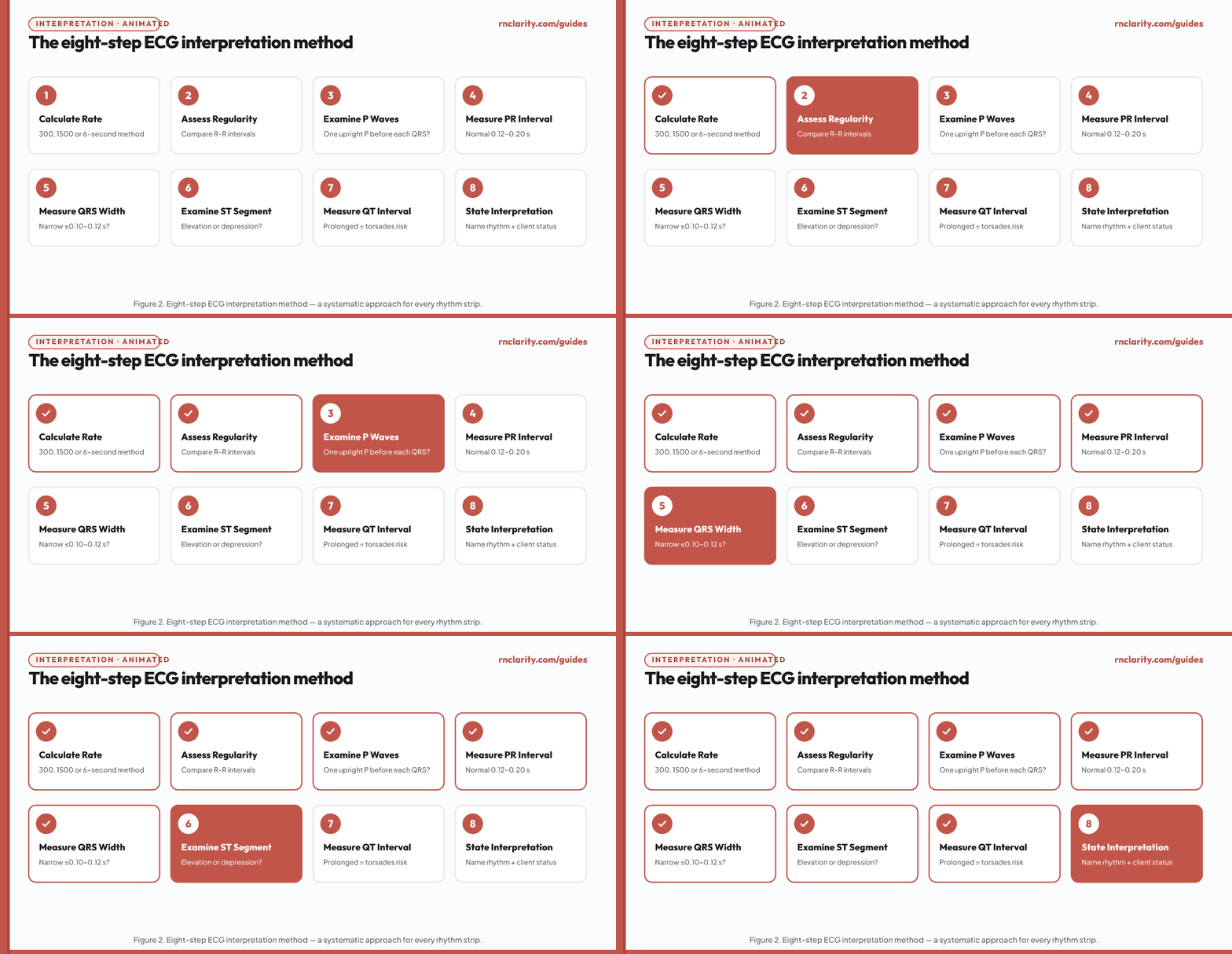
| Step | What to assess | Normal finding |
|---|---|---|
| 1 | Rate | 60–100 beats per minute |
| 2 | Regularity | Regular (R-R intervals equal) |
| 3 | P waves | Upright in lead II, one P before each QRS, consistent morphology |
| 4 | PR interval | 0.12–0.20 sec (3–5 small squares) |
| 5 | QRS width | ≤0.10 sec (≤2.5 small squares) — sometimes stated as <0.12 sec |
| 6 | ST segment | At isoelectric baseline — no elevation or depression |
| 7 | QT interval | 0.36–0.44 sec; corrected QTc ≤0.44 sec (men), ≤0.46 sec (women) |
| 8 | Interpretation | State the rhythm and any abnormalities |
Step-by-step application:
Step 1 — Rate: Use one of the three calculation methods described in section 5 below. State the rate in beats per minute.
Step 2 — Regularity: Measure the distance between consecutive R peaks (R-R interval) using calipers or the paper edge. If all R-R intervals are equal (within 0.04 seconds), the rhythm is regular. If they vary, determine whether the variation follows a pattern (regularly irregular) or is entirely random (irregularly irregular).
Step 3 — P waves: In lead II (the most common monitoring lead), normal P waves are upright, rounded, and identical. Ask: Is there a P wave before every QRS? Is there a QRS after every P wave? Are all P waves identical?
Step 4 — PR interval: Measure from the beginning of the P wave to the beginning of the QRS complex. A PR interval greater than 0.20 seconds indicates delayed AV conduction (first-degree AV block or longer). A PR that varies in length is a clue to second-degree AV block type I (Wenckebach).
Step 5 — QRS width: Measure from the first deflection of the QRS to the end of the last deflection, before the ST segment begins. A QRS wider than 0.10–0.12 seconds indicates either a bundle branch block or a ventricular origin of the impulse.
Step 6 — ST segment: The ST segment from the J point to the T wave should be isoelectric. Elevation or depression of ≥1 mm is clinically significant and must be reported immediately.
Step 7 — QT interval: Measure from the beginning of the QRS to the end of the T wave. A prolonged QT increases risk of torsades de pointes. The QT interval is rate-dependent; the corrected QTc is used for clinical decisions.
Step 8 — Interpretation: Synthesize all findings and state the rhythm interpretation, any abnormalities found, and the client's hemodynamic status.
Go deeper: For how clinical judgment applies to connecting ECG findings to priority nursing actions in NGN case studies, see the NGN case studies guide.
5. Rate Calculation Methods
Three methods are used in clinical practice. All are appropriate for NCLEX-RN® purposes; choose based on whether the rhythm is regular or irregular.
5.1 The 300-method (regular rhythms)
Count the number of large squares between two consecutive R waves. Divide 300 by that number.
Rate = 300 ÷ number of large squares between R peaks
Landmark table for quick estimation:
| Large squares between R peaks | Approximate rate |
|---|---|
| 1 | 300/min |
| 2 | 150/min |
| 3 | 100/min |
| 4 | 75/min |
| 5 | 60/min |
| 6 | 50/min |
Memorize: 300–150–100–75–60–50. When the R peak falls between large squares, interpolate.
5.2 The 1500-method (precise, regular rhythms)
Count the number of small squares between two R peaks. Divide 1500 by that number.
Rate = 1500 ÷ number of small squares between R peaks
5.3 The 6-second strip method (irregular rhythms)
Count the number of complete QRS complexes in a 6-second strip (30 large squares). Multiply by 10.
Rate = QRS complexes in 6 seconds × 10
This method estimates rate for irregular rhythms such as atrial fibrillation, where R-R intervals vary beat to beat.
6. Waves, Intervals and Segments
Precise measurement of ECG waveforms is essential for identifying dysrhythmias and drug toxicity. Every component of the normal ECG has a defined normal range.
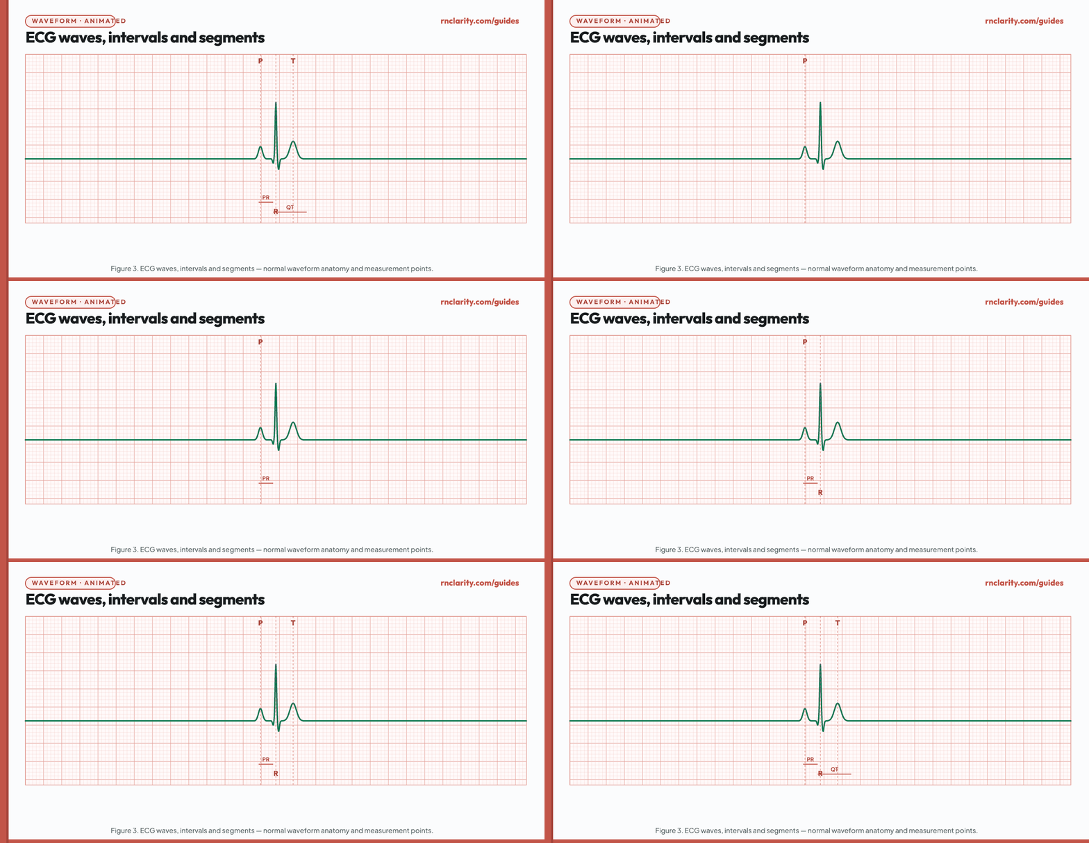
| Waveform or interval | Represents | Normal value |
|---|---|---|
| P wave | Atrial depolarization | Duration < 0.12 sec; amplitude < 2.5 mm in lead II |
| PR interval | Atrial depolarization + AV node delay | 0.12–0.20 sec |
| QRS complex | Ventricular depolarization | Duration ≤ 0.10–0.12 sec |
| ST segment | Period between ventricular depolarization and repolarization | Isoelectric; ≤ 1 mm deviation |
| T wave | Ventricular repolarization | Upright in most leads; peaked T waves suggest hyperkalemia |
| QT interval | Total ventricular electrical cycle | 0.36–0.44 sec; rate-corrected QTc ≤ 0.44 sec (men), ≤ 0.46 sec (women) |
| U wave | Slow repolarization of Purkinje fibers | Small positive deflection after T wave; prominent in hypokalemia |
Clinically significant abnormalities to know:
- Delta wave — slurred upstroke of the QRS in Wolff-Parkinson-White syndrome; indicates accessory pathway.
- Peaked (tall, narrow) T waves — hyperkalemia; the first and most specific ECG sign of elevated potassium.
- Flattened T waves with prominent U wave — hypokalemia.
- Prolonged QT — hypokalemia, hypomagnesemia, hypocalcemia, antiarrhythmic drugs (class Ia, Ic, III), antibiotics (macrolides, fluoroquinolones), antipsychotics, antidepressants.
- ST elevation — myocardial infarction (STEMI), pericarditis, Prinzmetal angina; requires immediate provider notification.
- ST depression — myocardial ischemia, digitalis effect, posterior MI.
7. Lead Placement, Telemetry Monitoring and Artifact
7.1 Lead placement
A standard 12-lead ECG uses 10 electrodes (4 limb, 6 precordial) to generate 12 views of cardiac electrical activity. Correct electrode placement is critical — misplacement as small as one intercostal space can alter QRS morphology and mimic bundle branch block or ST changes.
Limb electrode placement:
- RA — right arm, above wrist
- LA — left arm, above wrist
- RL — right leg, above ankle (ground electrode)
- LL — left leg, above ankle
Precordial electrode placement (V leads):
- V1 — 4th intercostal space, right sternal border
- V2 — 4th intercostal space, left sternal border
- V3 — midway between V2 and V4
- V4 — 5th intercostal space, midclavicular line
- V5 — anterior axillary line, same level as V4
- V6 — midaxillary line, same level as V4 and V5
Common misplacement errors:
- Reversing limb leads (RA-LA reversal produces inverted P, QRS, T in lead I and inverted P in aVL, mimicking right-sided placement or dextrocardia).
- Placing precordial leads too high or too low (mimics anterior ST changes or bundle branch block).
7.2 Telemetry monitoring
Continuous telemetry is used in medical-surgical, step-down, and intensive care settings to detect dysrhythmias in real time. Lead II is the most commonly used monitoring lead because P waves are typically most visible and upright in that view.
Three-lead telemetry monitors typically display Leads I, II, and III (or a modified chest lead MCL1). Five-lead systems add leads V1 through V6 selectively and are preferred for identifying bundle branch blocks and differentiating wide-complex tachycardias.
Nursing responsibilities for telemetry monitoring include:
- Confirming electrode adherence at each shift; replacing dried or detached electrodes promptly.
- Documenting a rhythm strip and client assessment at the beginning of each shift and with any rhythm change.
- Recognizing alarm fatigue risk — do not silence alarms without assessing the client.
- Correlating any rhythm change with the client's clinical status before notifying the provider.
7.3 Artifact identification
Artifact is any waveform on the ECG that does not represent true cardiac electrical activity. Failing to recognize artifact can lead to inappropriate treatment.
| Artifact type | Appearance | Common cause |
|---|---|---|
| Muscle artifact (somatic tremor) | Irregular, fuzzy baseline; R-R intervals may appear irregular | Shivering, Parkinson disease, patient movement |
| AC interference (60-Hz) | Fine, regular sawtooth baseline interference | Poor grounding; electrical equipment near client |
| Wandering baseline | Gradual drift of baseline up and down | Poor electrode contact, patient breathing, diaphoresis |
| Motion artifact | Erratic, large deflections obscuring true rhythm | Patient movement; poor lead adherence |
The nursing priority when artifact appears is to assess the client before treating the tracing. A monitor that shows a flat line while the client is awake, alert, and talking is artifact, not asystole.
Go deeper: For how pharmacological agents such as antiarrhythmics affect conduction and create ECG changes, see the pharmacology guide.
8. Sinus Rhythms
Sinus rhythms originate in the SA node. All sinus rhythms share three features: upright P waves in lead II with consistent morphology, one P wave before each QRS, and a PR interval of 0.12–0.20 seconds.
8.1 Normal sinus rhythm
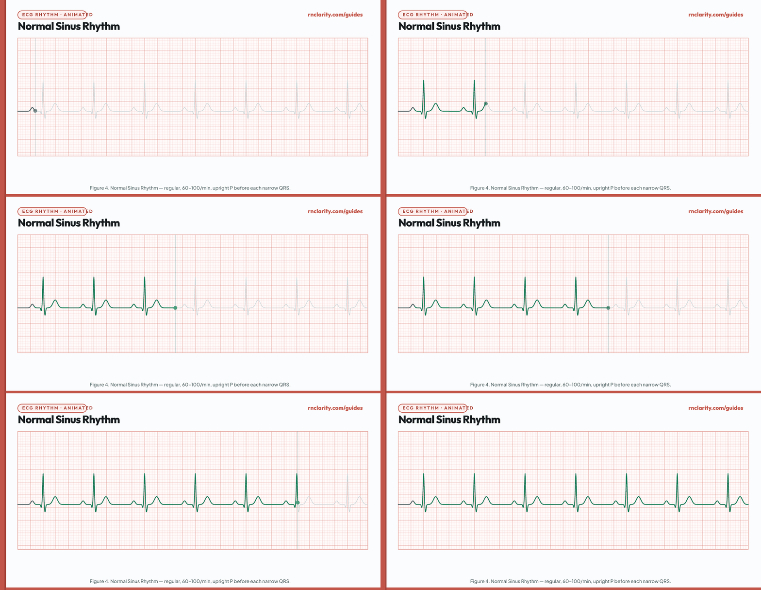
| Feature | Normal sinus rhythm finding |
|---|---|
| Rate | 60–100/min |
| Regularity | Regular |
| P waves | Upright in lead II; one per QRS; consistent |
| PR interval | 0.12–0.20 sec |
| QRS | ≤ 0.10–0.12 sec; narrow |
| Nursing priority | No dysrhythmia; continue routine monitoring |
Normal sinus rhythm is the baseline against which all other rhythms are compared. Every step of the eight-step method yields a normal finding.
8.2 Sinus bradycardia
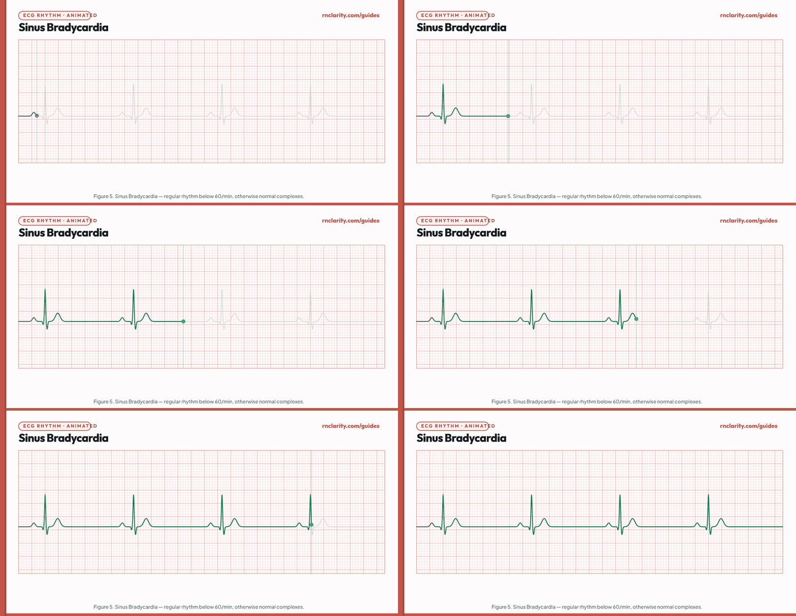
| Feature | Sinus bradycardia finding |
|---|---|
| Rate | < 60/min |
| Regularity | Regular |
| P waves | Upright, one per QRS |
| PR interval | 0.12–0.20 sec |
| QRS | Narrow |
| Common causes | Athletes (normal), vagal stimulation, beta-blockers, calcium channel blockers, digoxin, hypothyroidism, increased intracranial pressure (Cushing's triad), SA node disease |
Nursing priority: Assess hemodynamic tolerance. A resting bradycardia of 50/min in an athletic client with no symptoms may need observation rather than acute treatment. Bradycardia with cardiopulmonary compromise (such as hypotension, acutely altered mental status, shock, ischemic chest discomfort, or acute heart failure) requires an immediate response. The 2025 AHA adult bradycardia algorithm gives atropine 1 mg IV bolus as the initial dose. If atropine is ineffective, prepare transcutaneous pacing and/or a dopamine or epinephrine infusion according to the resuscitation protocol while addressing reversible causes.
NCLEX-RN® trap: Do not automatically treat every bradycardia. An athlete with a resting heart rate of 48/min, normal blood pressure, and no symptoms requires monitoring, not intervention.
8.3 Sinus tachycardia
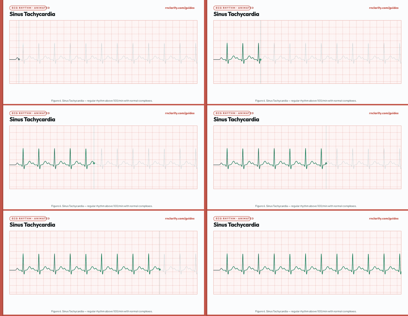
| Feature | Sinus tachycardia finding |
|---|---|
| Rate | 100–150/min (sinus maximum is approximately 220 minus age) |
| Regularity | Regular |
| P waves | Upright, one per QRS; may merge with preceding T wave at very fast rates |
| PR interval | 0.12–0.20 sec or slightly shortened |
| QRS | Narrow |
| Common causes | Pain, anxiety, fever, hypovolemia, anemia, hypoxia, hyperthyroidism, pulmonary embolism, heart failure, stimulants, caffeine |
Nursing priority: Sinus tachycardia is nearly always a compensatory response to an underlying condition. Identify and treat the cause — do not treat the tachycardia alone. Administer antipyretics for fever, analgesics for pain, IV fluids for hypovolemia. A sudden-onset tachycardia with hemodynamic compromise requires urgent provider notification.
8.4 Sinus arrhythmia
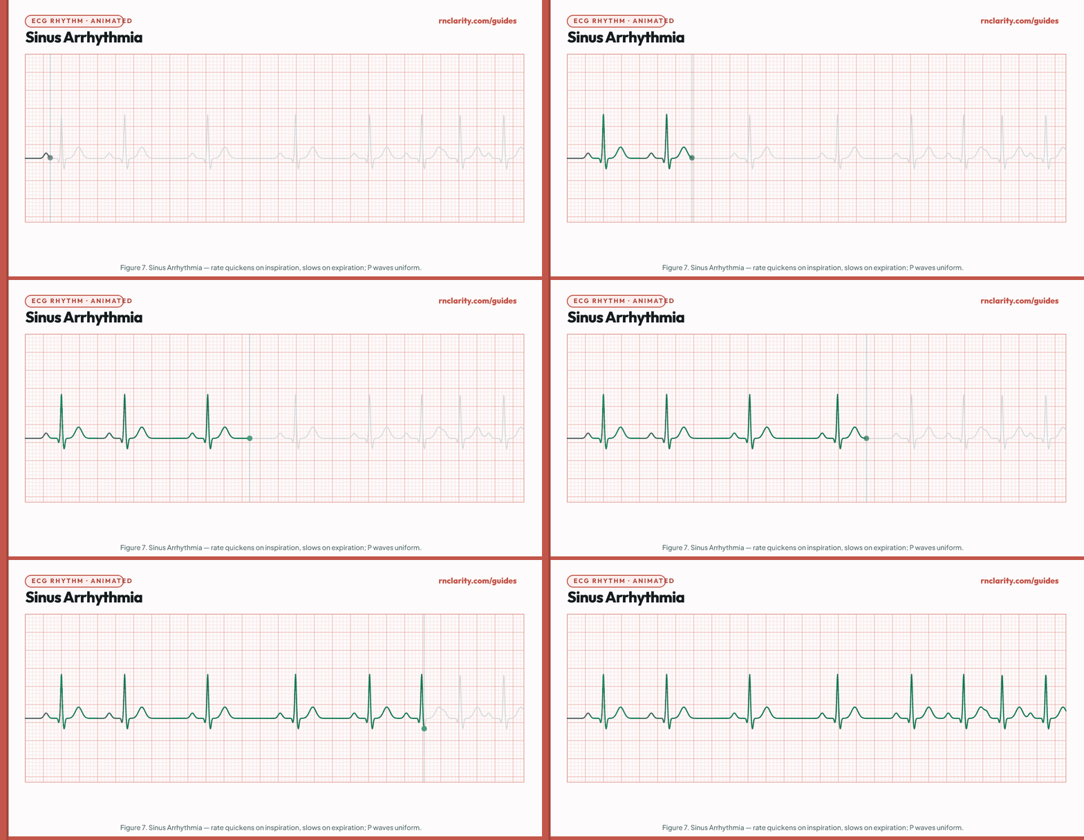
Sinus arrhythmia is a normal variant in which the SA node fires slightly faster during inspiration and slower during expiration. R-R intervals vary but the variation follows the respiratory cycle. P waves are upright and uniform. It is most common in children and young adults and requires no treatment.
Key distinction from pathological irregular rhythms: In sinus arrhythmia, the variation in R-R intervals follows a rhythmic pattern tied to breathing and all P-wave morphologies are identical. Atrial fibrillation is irregularly irregular with no true P waves.
9. Atrial Rhythms
Atrial rhythms originate in ectopic foci within the atrial tissue, outside the SA node. Because the impulse still travels the normal His-Purkinje route, QRS complexes are typically narrow, but P-wave morphology differs from sinus P waves.
9.1 Premature atrial complex
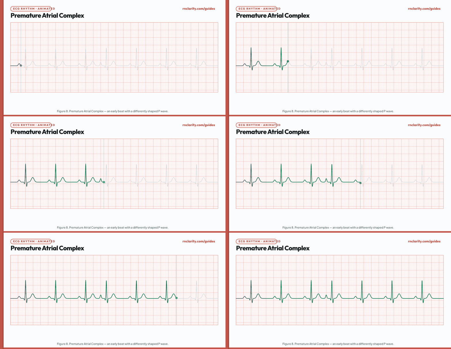
A premature atrial complex (PAC) occurs when an ectopic atrial focus fires before the next expected sinus impulse. The P wave is early and has a different morphology than the sinus P waves. The QRS is narrow (the ventricles conduct normally). A non-compensatory pause usually follows.
| Feature | PAC finding |
|---|---|
| Rate | Underlying sinus rate; interrupted by early beat |
| Regularity | Irregular (early beat breaks the pattern) |
| P wave of PAC | Early, different morphology; may be inverted, notched, or hidden in the preceding T wave |
| PR interval (PAC) | May be longer or shorter than sinus PR |
| QRS | Narrow; same as sinus beats |
| Common causes | Caffeine, alcohol, tobacco, electrolyte disturbance, stress, atrial enlargement, stimulant medications |
Nursing priority: Isolated PACs are common and often benign. Frequent PACs (> 6/min) or PAC runs may precede atrial fibrillation or flutter and warrant provider notification. Eliminate triggers (caffeine, tobacco, stimulants).
9.2 Atrial flutter
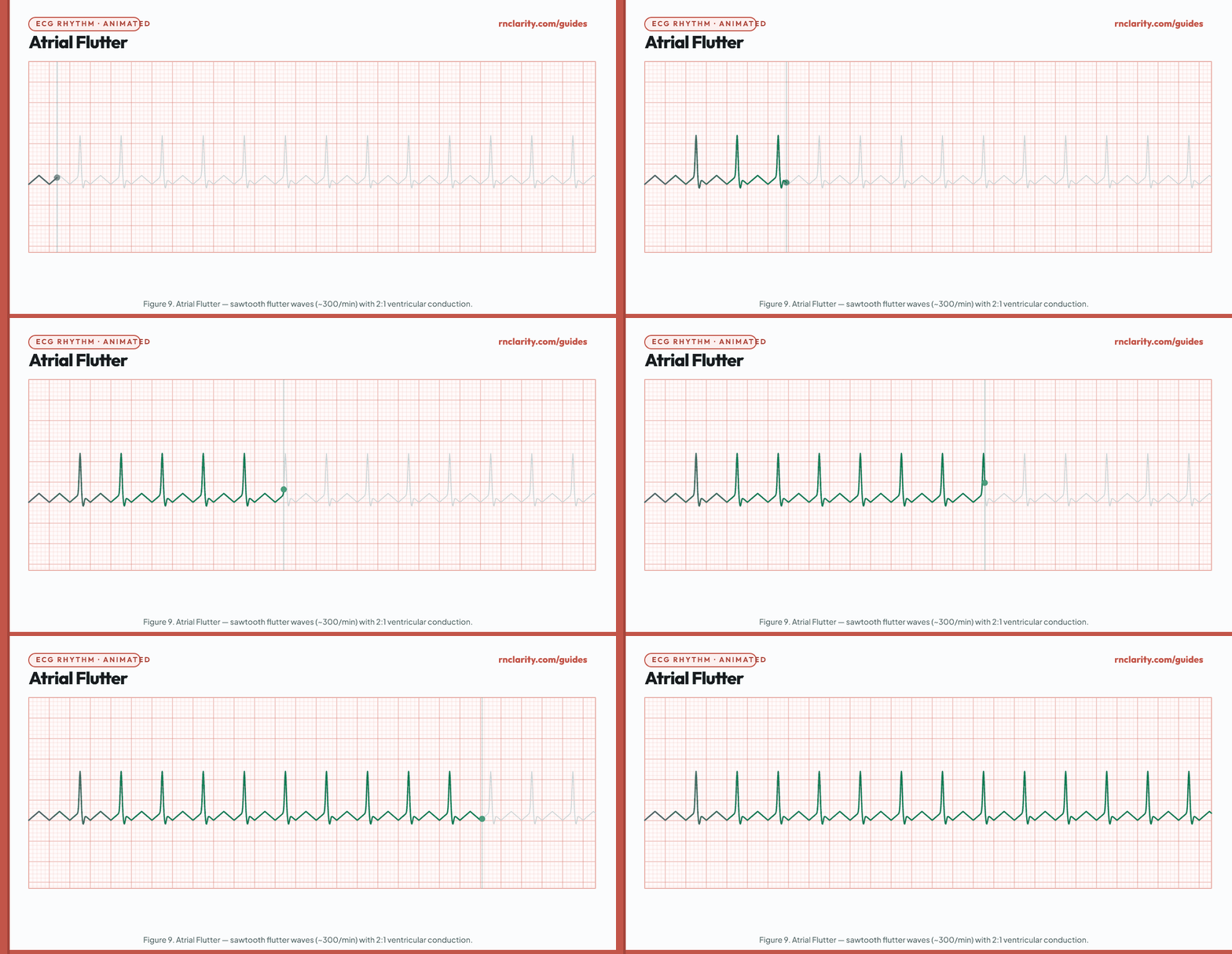
| Feature | Atrial flutter finding |
|---|---|
| Atrial rate | 250–350/min (typically ~300/min) |
| Ventricular rate | Depends on AV conduction ratio: 2:1 = ~150/min; 4:1 = ~75/min |
| Regularity | Ventricular rhythm is regularly regular (fixed ratio) or regularly irregular (variable ratio) |
| P waves | Replaced by sawtooth flutter waves (F waves); no isoelectric baseline between waves |
| QRS | Narrow; flutter waves may hide within QRS |
| Common causes | Structural heart disease, post-cardiac surgery, hyperthyroidism, COPD, pulmonary embolism |
Nursing priority: Atrial flutter carries stroke risk due to impaired atrial contractility and potential thrombus formation. Immediate assessment includes heart rate, blood pressure, and oxygen saturation. Unstable flutter (hypotension, chest pain, syncope) requires immediate synchronized cardioversion. Stable flutter is managed with rate control (beta-blockers, calcium channel blockers) and anticoagulation. Notify the provider promptly.
NCLEX-RN® key fact: A ventricular rate of exactly 150/min in a client with a narrow-complex tachycardia should prompt suspicion of atrial flutter with 2:1 conduction. Look carefully for flutter waves at twice that rate in the isoelectric segments.
9.3 Atrial fibrillation
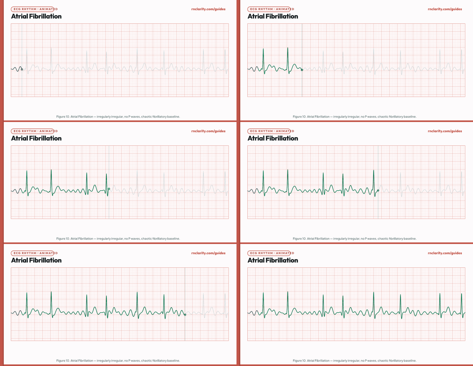
| Feature | Atrial fibrillation finding |
|---|---|
| Atrial activity | 350–600 chaotic impulses per minute; no organized P waves; fibrillatory (f) waves |
| Ventricular rate | Controlled: 60–100/min; rapid: > 100/min; slow: < 60/min (may indicate AV block or digoxin toxicity) |
| Regularity | Irregularly irregular — no two consecutive R-R intervals are the same |
| P waves | Absent; replaced by chaotic fibrillatory baseline |
| QRS | Narrow (unless aberrant conduction or bundle branch block) |
| Common causes | Hypertension, heart failure, valvular disease, coronary artery disease, hyperthyroidism, alcohol ("holiday heart"), post-cardiac surgery, COPD |
Nursing priority: Atrial fibrillation is the most common sustained arrhythmia encountered in clinical practice. Key nursing priorities:
- Assess hemodynamic status (blood pressure, pulse, level of consciousness).
- Unstable AF (hypotension, chest pain, pulmonary edema) → immediate synchronized cardioversion.
- Stable AF with rapid ventricular response → rate control medications (beta-blockers: metoprolol; non-dihydropyridine calcium channel blockers: diltiazem, verapamil; digoxin for low blood pressure).
- Anticoagulation is a priority — AF with > 48 hours duration or unknown onset requires anticoagulation before cardioversion to prevent stroke from atrial thrombus.
- Monitor for signs of stroke: facial droop, arm drift, speech changes (FAST assessment).
NCLEX-RN® high-yield: The signature finding of atrial fibrillation is an irregularly irregular ventricular rhythm with no identifiable P waves. Both features must be present to identify AF.
9.4 Supraventricular tachycardia
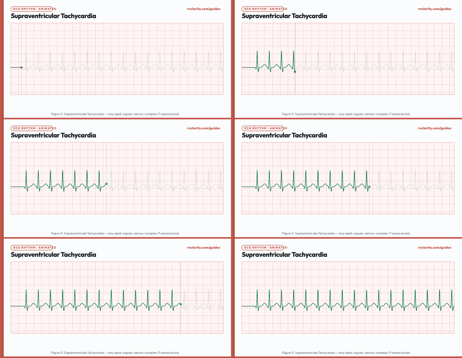
| Feature | SVT finding |
|---|---|
| Rate | 150–250/min |
| Regularity | Regular (very) |
| P waves | Not visible (buried in QRS or preceding T wave) |
| QRS | Narrow (< 0.12 sec); identical beats |
| Common causes | Reentry involving the AV node (AVNRT most common); accessory pathway (AVRT); emotional stress, caffeine, stimulants, thyroid disease |
Nursing priority: Many SVT episodes terminate spontaneously or with vagal maneuvers. For a stable client, the nurse may assist with the Valsalva maneuver (straining, bearing down) or carotid sinus massage (physician-performed). If vagal maneuvers fail, adenosine 6 mg IV rapid push followed by a saline flush is the pharmacological first-line treatment per ACLS — it must be pushed rapidly into a large peripheral or central IV closest to the heart. For unstable SVT (hemodynamic compromise), synchronized cardioversion is indicated.
Go deeper: For clinical judgment frameworks applied to tachyarrhythmia prioritization scenarios, see the practice questions guide.
10. Junctional Rhythms
Junctional rhythms originate in the AV node or bundle of His. Because the impulse simultaneously activates the atria (retrograde) and ventricles (antegrade), P waves are inverted (retrograde), occur just before, within, or just after the QRS, or are absent.
10.1 Junctional escape rhythm
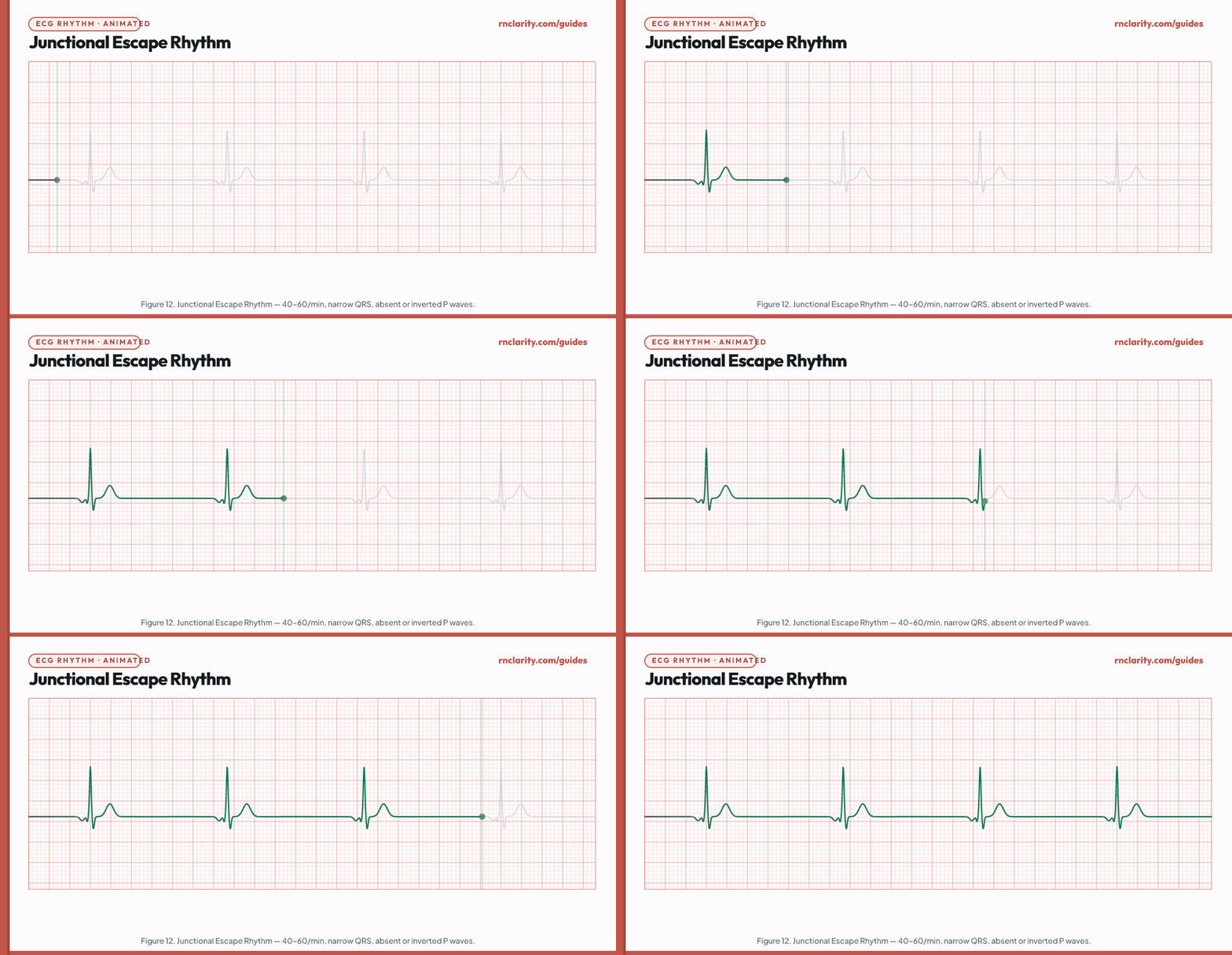
| Feature | Junctional escape rhythm finding |
|---|---|
| Rate | 40–60/min |
| Regularity | Regular |
| P waves | Inverted in lead II; may appear before QRS (short PR ≤ 0.12 sec), within QRS (not visible), or after QRS (RP interval); or absent |
| QRS | Narrow (≤ 0.12 sec) |
| Common causes | SA node failure, high-degree AV block, inferior MI, vagal stimulation, digoxin toxicity |
Nursing priority: Junctional escape rhythm is a protective mechanism — the AV node has taken over because the SA node has failed or because AV conduction is blocked. Assess the client for hemodynamic tolerance. A rate of 50/min with stable blood pressure and normal mentation may be observed while the cause is sought. Symptomatic junctional bradycardia is treated with atropine; pacing may be required.
10.2 Accelerated junctional rhythm
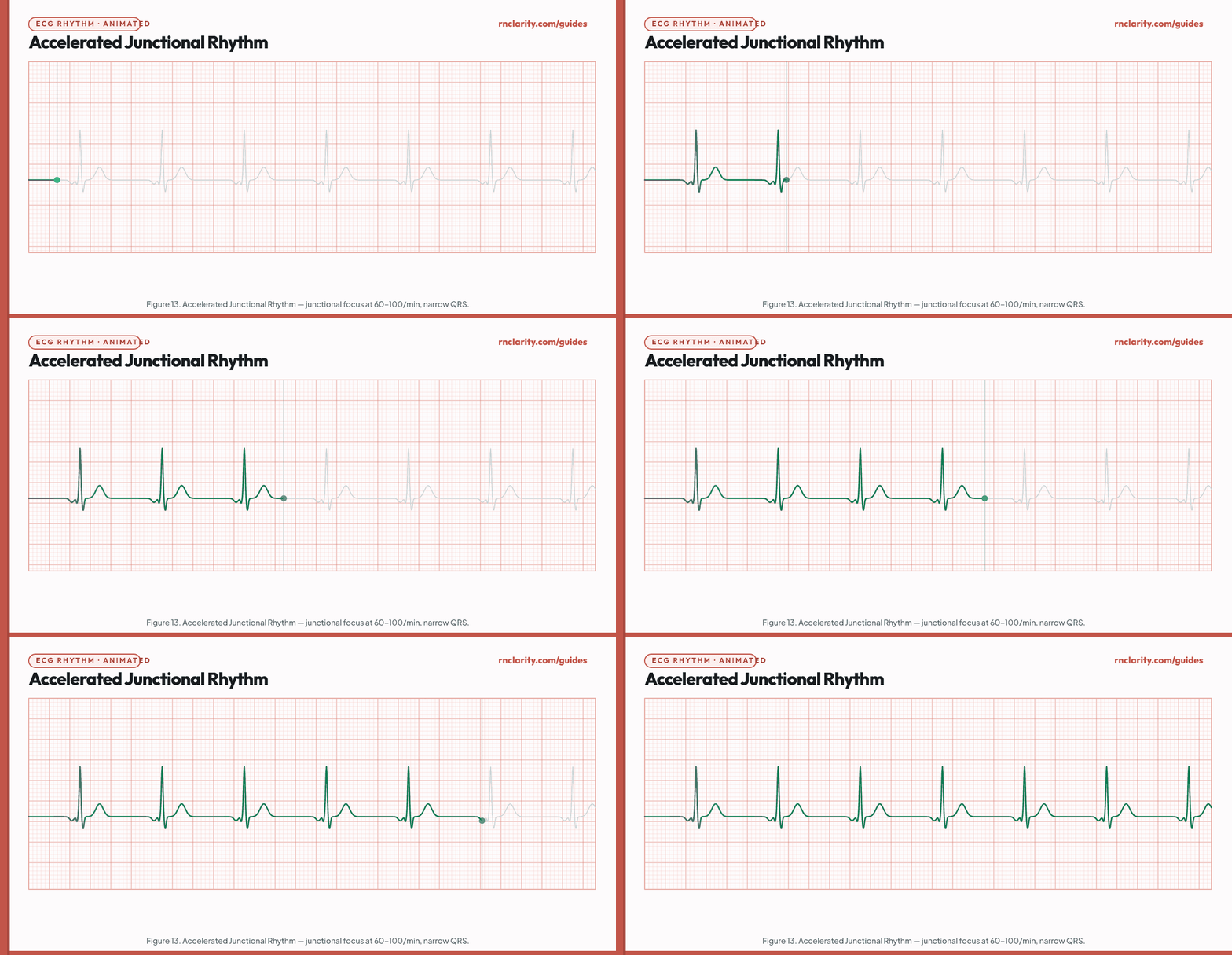
When a junctional focus fires faster than its normal escape rate (60–100/min), the rhythm is called accelerated junctional rhythm. It may compete with the SA node, producing isorhythmic AV dissociation. Common causes include digoxin toxicity, inferior MI, and rheumatic heart disease. Assess the client and notify the provider; if digoxin toxicity is suspected, check the serum digoxin level.
11. AV Blocks
AV blocks represent impaired conduction through the AV node or bundle of His. They are classified by degree (first, second, or third) based on the severity of conduction impairment.
11.1 First-degree AV block
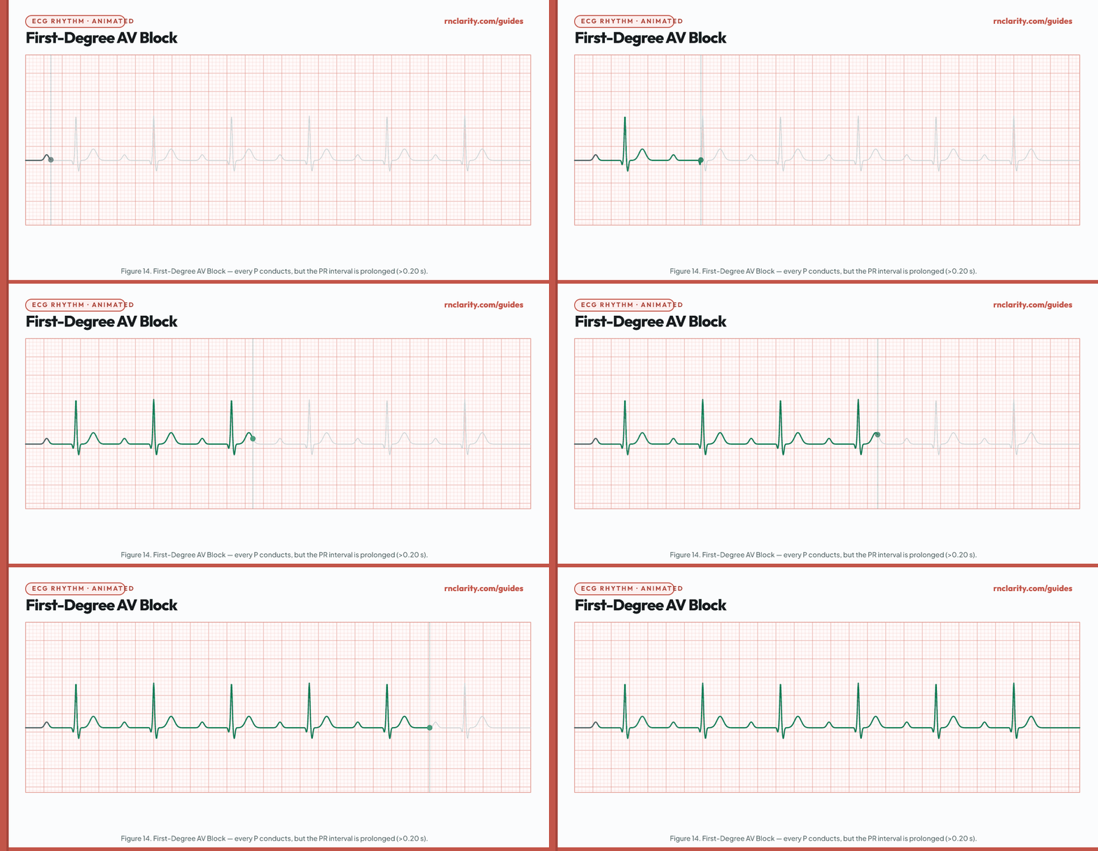
| Feature | First-degree AV block finding |
|---|---|
| Rate | Underlying sinus rate (usually 60–100/min) |
| Regularity | Regular |
| P waves | Upright in lead II; one before each QRS |
| PR interval | > 0.20 sec (more than one large square); consistent from beat to beat |
| QRS | Narrow |
| Clinical significance | Every impulse still conducts; the AV node is simply slower than normal |
| Common causes | Inferior MI, beta-blockers, calcium channel blockers, digoxin, aging, hypokalemia, Lyme disease |
Nursing priority: First-degree AV block alone does not require treatment. Document it, identify potential causes (especially if the client is on AV-slowing medications), and monitor for progression to higher-degree block. Notify the provider when a new first-degree block is identified.
11.2 Second-degree AV block type I (Wenckebach / Mobitz I)
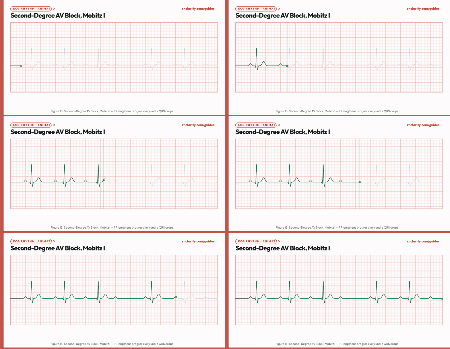
| Feature | Mobitz I finding |
|---|---|
| Rate | Atrial rate regular; ventricular rate slightly irregular (dropped beats) |
| P waves | Regular; more P waves than QRS complexes |
| PR interval | Progressively lengthens until one P wave is not followed by a QRS (the QRS is "dropped"); then the cycle resets |
| QRS | Narrow when conducted |
| R-R pattern | Gradually shortens before the dropped beat; longest R-R interval is less than twice the shortest |
| Common causes | Inferior MI (right coronary artery supplies AV node), increased vagal tone, inferior ischemia, beta-blockers, digoxin |
Nursing priority: Wenckebach is often transient and hemodynamically tolerated. Assess the client for symptoms (dizziness, near-syncope). Notify the provider. Monitor closely for progression to higher-degree block, particularly in the setting of inferior MI.
Memory trick: "Longer, longer, longer, DROP — then you have a Wenckebach."
11.3 Second-degree AV block type II (Mobitz II)
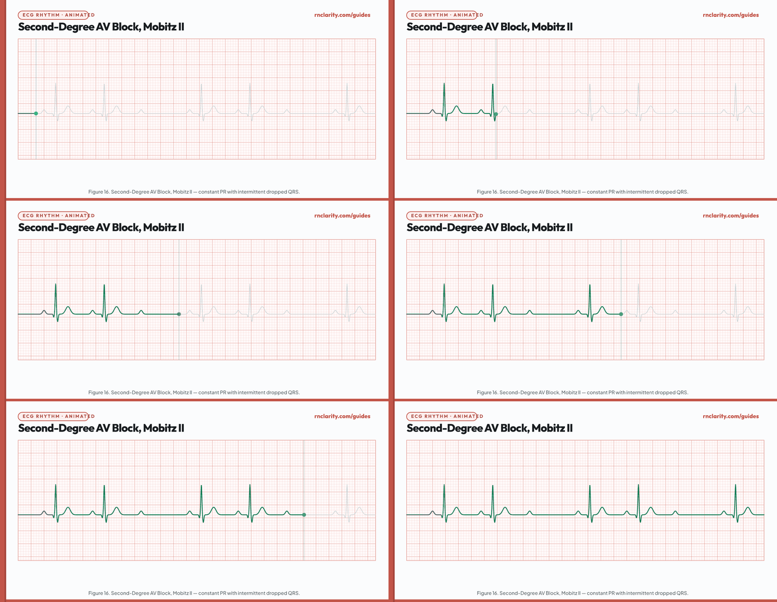
| Feature | Mobitz II finding |
|---|---|
| Rate | Atrial rate regular; ventricular rate depends on block ratio (2:1, 3:1, etc.) |
| P waves | Regular; more P waves than QRS complexes |
| PR interval | Constant on conducted beats; then a P wave suddenly drops without prior PR prolongation |
| QRS | Often wide (bundle branch block pattern) — block is below the AV node, in the bundle of His or bundle branches |
| Clinical significance | Potentially life-threatening — can progress suddenly to complete (third-degree) AV block without warning |
| Common causes | Anterior MI, calcific disease of the conduction system, Lyme disease |
Nursing priority: Mobitz II is a serious finding requiring immediate provider notification. Transcutaneous pacing equipment should be prepared and placed on standby. The client should not ambulate unattended because sudden progression to complete AV block can cause hemodynamic collapse. A permanent pacemaker is typically required.
Critical distinction — Mobitz I vs. Mobitz II:
| Feature | Mobitz I (Wenckebach) | Mobitz II |
|---|---|---|
| PR interval | Progressively lengthens before drop | Constant before drop |
| QRS width | Usually narrow | Often wide |
| Location of block | AV node | Bundle of His / bundle branches |
| Risk of progression | Lower | Higher — may go to complete AV block suddenly |
| Common cause | Inferior MI | Anterior MI |
11.4 Third-degree (complete) AV block
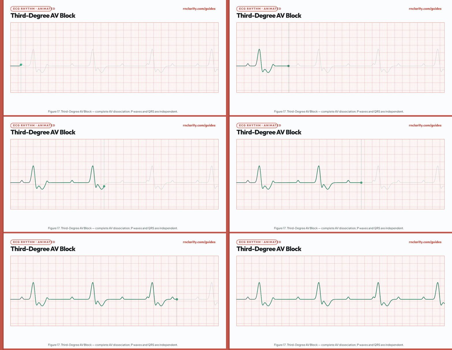
| Feature | Third-degree AV block finding |
|---|---|
| Atrial rate | 60–100/min (SA node fires normally) |
| Ventricular rate | 20–60/min (junctional escape 40–60; ventricular escape 20–40) |
| Regularity | Both atrial and ventricular rhythms are regular, but independent of each other |
| P waves | Regular; no fixed relationship to QRS |
| PR interval | No consistent PR interval — P waves march through QRS complexes |
| QRS | Narrow if junctional escape pacemaker; wide and bizarre if ventricular escape pacemaker |
| Clinical significance | Medical emergency — complete failure of AV conduction; atria and ventricles beat independently |
Nursing priority: Third-degree AV block requires emergency provider notification and preparation for immediate transcutaneous pacing. If the client is hemodynamically unstable (hypotension, altered mental status, chest pain), initiate transcutaneous pacing without delay. Atropine is less effective in complete heart block but may be attempted. Permanent pacemaker implantation is the definitive treatment.
Go deeper: For managing clients with cardiac emergencies in NGN case format, see the NGN case studies guide.
12. Ventricular Ectopy
Ventricular ectopic beats originate below the bundle of His, outside the normal conduction pathway. Because conduction spreads cell-to-cell through the ventricular myocardium rather than via the fast His-Purkinje system, QRS complexes are wide (> 0.12 sec) and have a bizarre, abnormal morphology. The T wave deflects in the opposite direction to the QRS (discordant).
12.1 Isolated premature ventricular complex
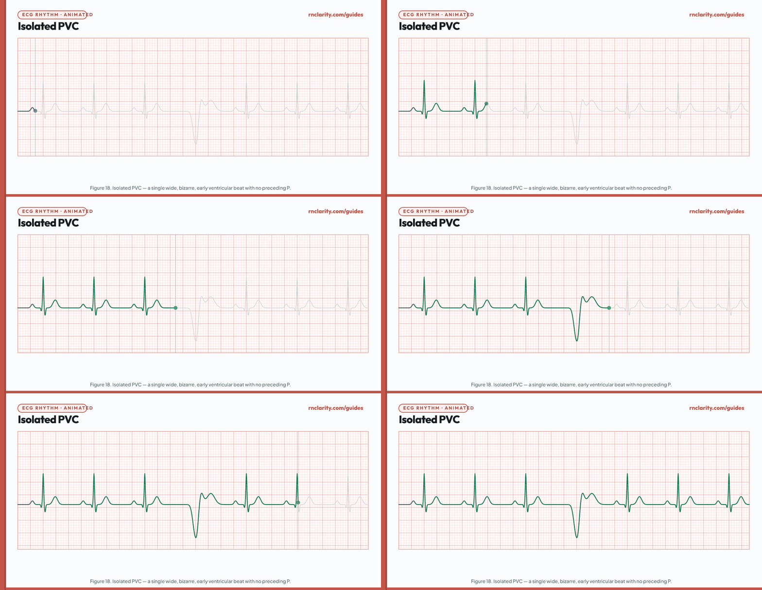
| Feature | Isolated PVC finding |
|---|---|
| Timing | Early — occurs before the next expected sinus beat |
| P wave | Absent before the PVC |
| QRS | Wide (> 0.12 sec), bizarre morphology |
| T wave | Opposite direction to QRS (discordant) |
| Pause | Full compensatory pause (the SA node fires on time; the total R-R interval containing the PVC = 2× normal R-R) |
| Common causes | Hypoxia, hypokalemia, hypomagnesemia, digoxin toxicity, caffeine, cocaine, myocardial ischemia/infarction, heart failure, cardiomyopathy |
Nursing priority: Isolated PVCs are common and occur in healthy individuals. In clients with cardiac disease, PVCs may indicate underlying ischemia or electrolyte imbalance. Assess for hypoxia, check electrolytes (potassium, magnesium), and review current medications (digoxin level if applicable). Notify the provider. Frequent PVCs (> 6/min), multifocal PVCs, R-on-T phenomenon (PVC falling on the T wave of the preceding beat), or runs of PVCs are higher-risk patterns warranting urgent notification.
12.2 Ventricular bigeminy
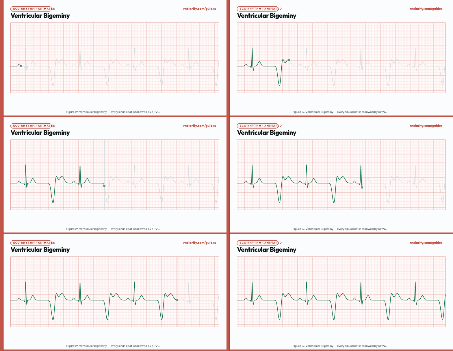
Ventricular bigeminy is a pattern in which every other beat is a PVC — one sinus beat, one PVC, one sinus beat, one PVC, repeating continuously. Trigeminy is every third beat a PVC. These patterns are associated with digoxin toxicity, hypokalemia, and structural heart disease.
Nursing priority: Assess client and check pulse rate, since PVCs may not perfuse adequately. Check digoxin level if applicable. Check serum potassium and magnesium. Notify the provider. If the effective ventricular rate is < 40/min (because the PVCs are not perfusing), treat as bradycardia.
12.3 Ventricular couplet
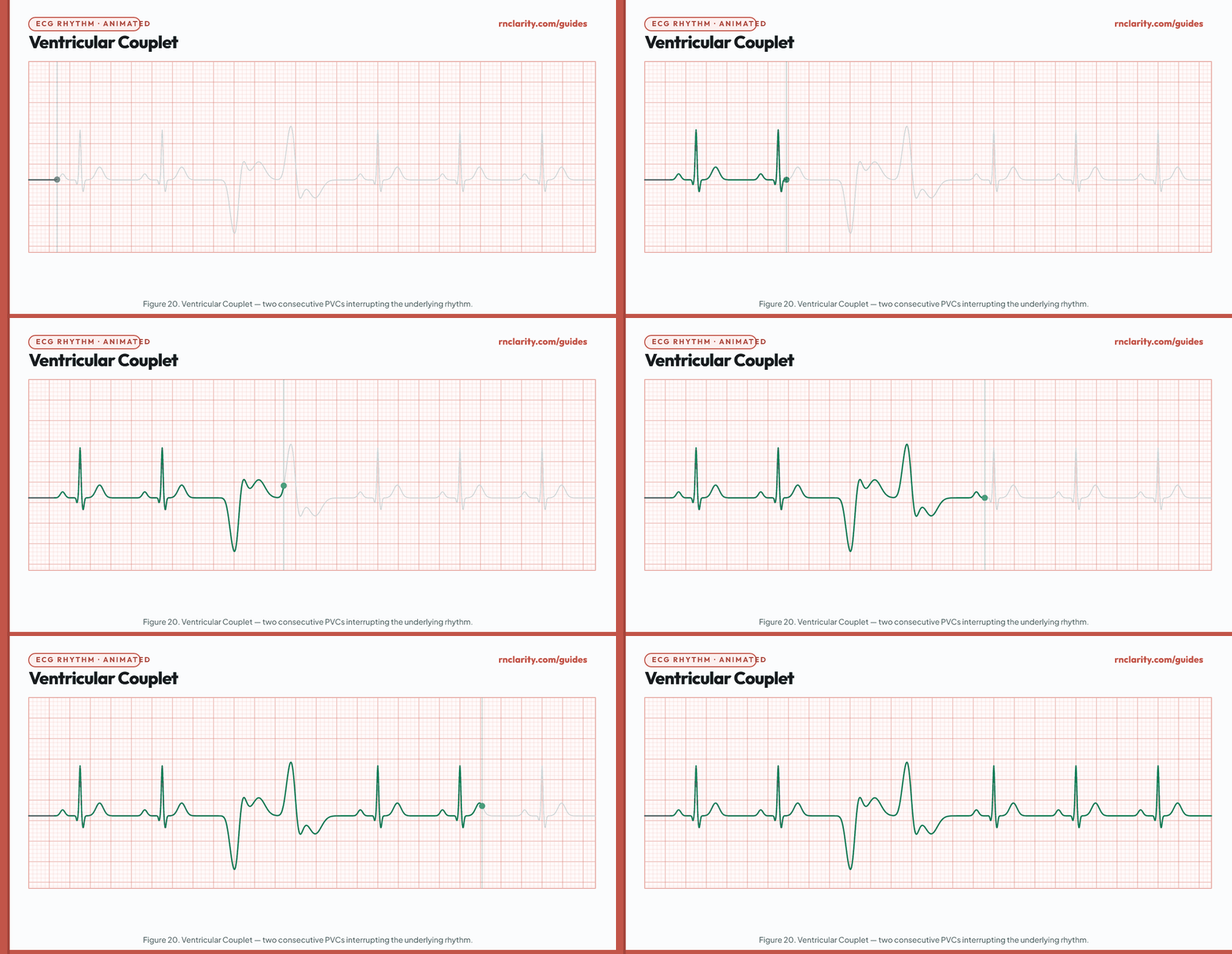
A ventricular couplet is two consecutive PVCs. Couplets indicate increased ventricular irritability. Three or more consecutive PVCs constitute a run of ventricular tachycardia. The presence of couplets, particularly in a client with known coronary artery disease or cardiomyopathy, requires prompt provider notification and consideration of electrolyte replacement and antiarrhythmic therapy.
12.4 Monomorphic ventricular tachycardia
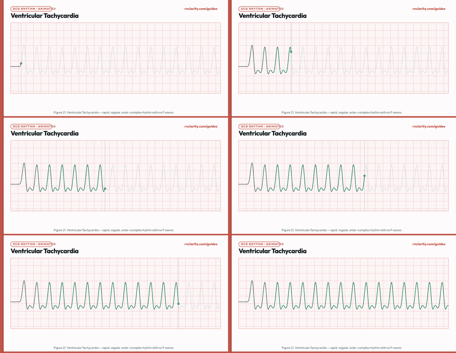
| Feature | Monomorphic VT finding |
|---|---|
| Rate | 100–250/min |
| Regularity | Regular or slightly irregular |
| P waves | Absent or dissociated (AV dissociation) |
| QRS | Wide (> 0.12 sec), bizarre, identical morphology |
| Clinical significance | Potentially life-threatening — may degenerate into ventricular fibrillation |
| Common causes | Myocardial infarction, cardiomyopathy, electrolyte disturbance, long QT syndrome, digoxin toxicity, cocaine |
Nursing priority:
- Pulseless VT: Treat as cardiac arrest — start CPR, call for help, defibrillate immediately (unsynchronized shock).
- Sustained VT with pulse, unstable (hypotension, chest pain, altered mental status): Synchronized cardioversion.
- Sustained VT with pulse, stable: Antiarrhythmic therapy (amiodarone 150 mg IV over 10 minutes is first-line per ACLS) and immediate provider/rapid response notification.
12.5 Torsades de pointes
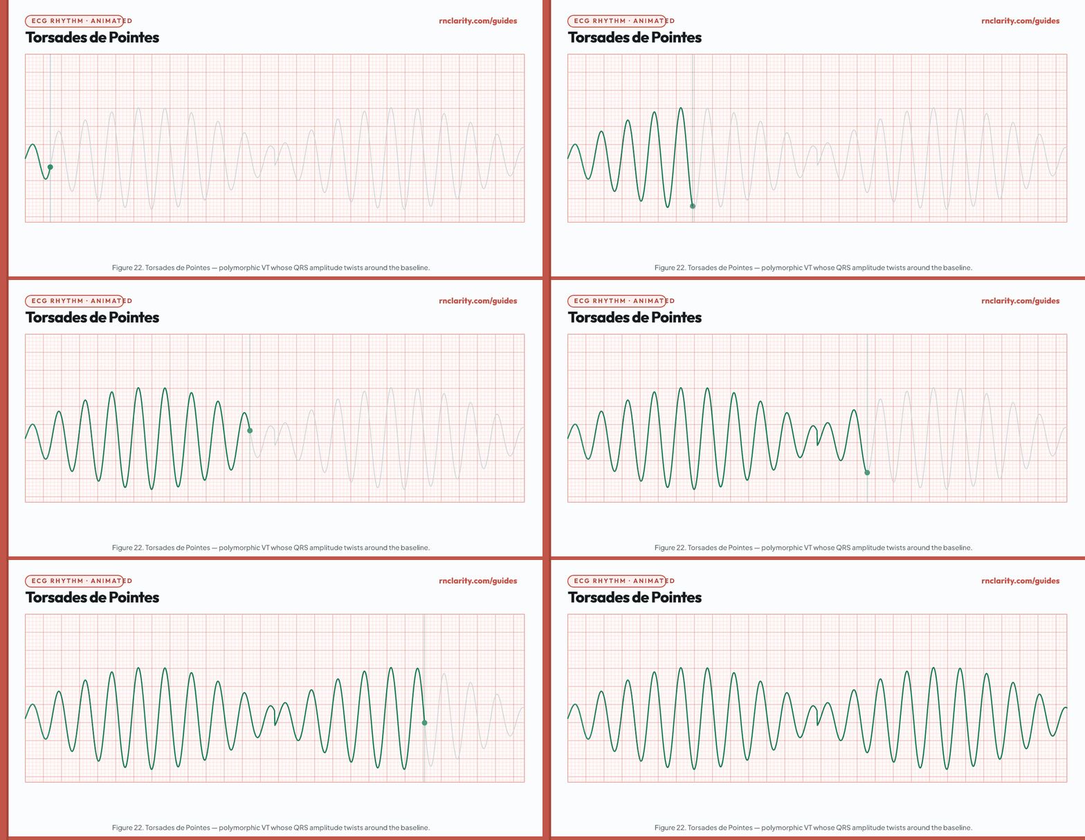
| Feature | Torsades de pointes finding |
|---|---|
| Rate | 150–300/min |
| Appearance | Polymorphic VT — QRS complexes twist around the isoelectric baseline, varying in amplitude and direction; the name means "twisting of the points" in French |
| Preceding rhythm | Prolonged QT interval on the preceding sinus beats is characteristic |
| Common causes | Hypokalemia, hypomagnesemia, hypocalcemia; QT-prolonging medications (class Ia and III antiarrhythmics, certain antibiotics, antipsychotics, antihistamines); congenital long QT syndrome |
Nursing priority: Magnesium sulfate 1–2 g IV over 5–20 minutes is the treatment of choice for torsades de pointes, regardless of the serum magnesium level. Correct electrolyte abnormalities (potassium, calcium). Identify and discontinue QT-prolonging medications. Unstable or pulseless torsades is treated with unsynchronized defibrillation. Prevention includes monitoring QTc in clients receiving QT-prolonging drugs.
NCLEX-RN® high-yield: Magnesium is the antidote for torsades de pointes. Always associate torsades with a prolonged QT and electrolyte imbalances (especially hypokalemia and hypomagnesemia).
13. Arrest Rhythms
Arrest rhythms are those that produce no effective cardiac output. Recognition must be immediate; survival depends on rapid initiation of CPR and defibrillation within minutes.
13.1 Ventricular fibrillation
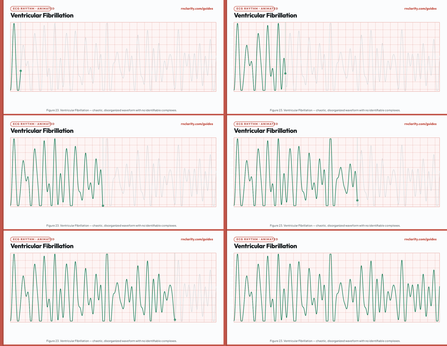
| Feature | VF finding |
|---|---|
| Rate | No organized rate — chaotic electrical activity |
| Rhythm | Completely chaotic |
| Waveforms | No identifiable P waves, QRS complexes, or T waves; undulating, irregular waveform |
| Coarse vs fine | Coarse VF has large, irregular deflections; fine VF has small ones. Coarse VF is more amenable to defibrillation |
| Clinical significance | No cardiac output — cardiac arrest |
Nursing priority: VF is treated with immediate unsynchronized defibrillation (200 J biphasic or 360 J monophasic for the first shock) combined with high-quality CPR. Epinephrine 1 mg IV/IO every 3–5 minutes and amiodarone 300 mg IV/IO bolus are administered after the initial defibrillation attempts. Never delay defibrillation to obtain IV access.
13.2 Asystole
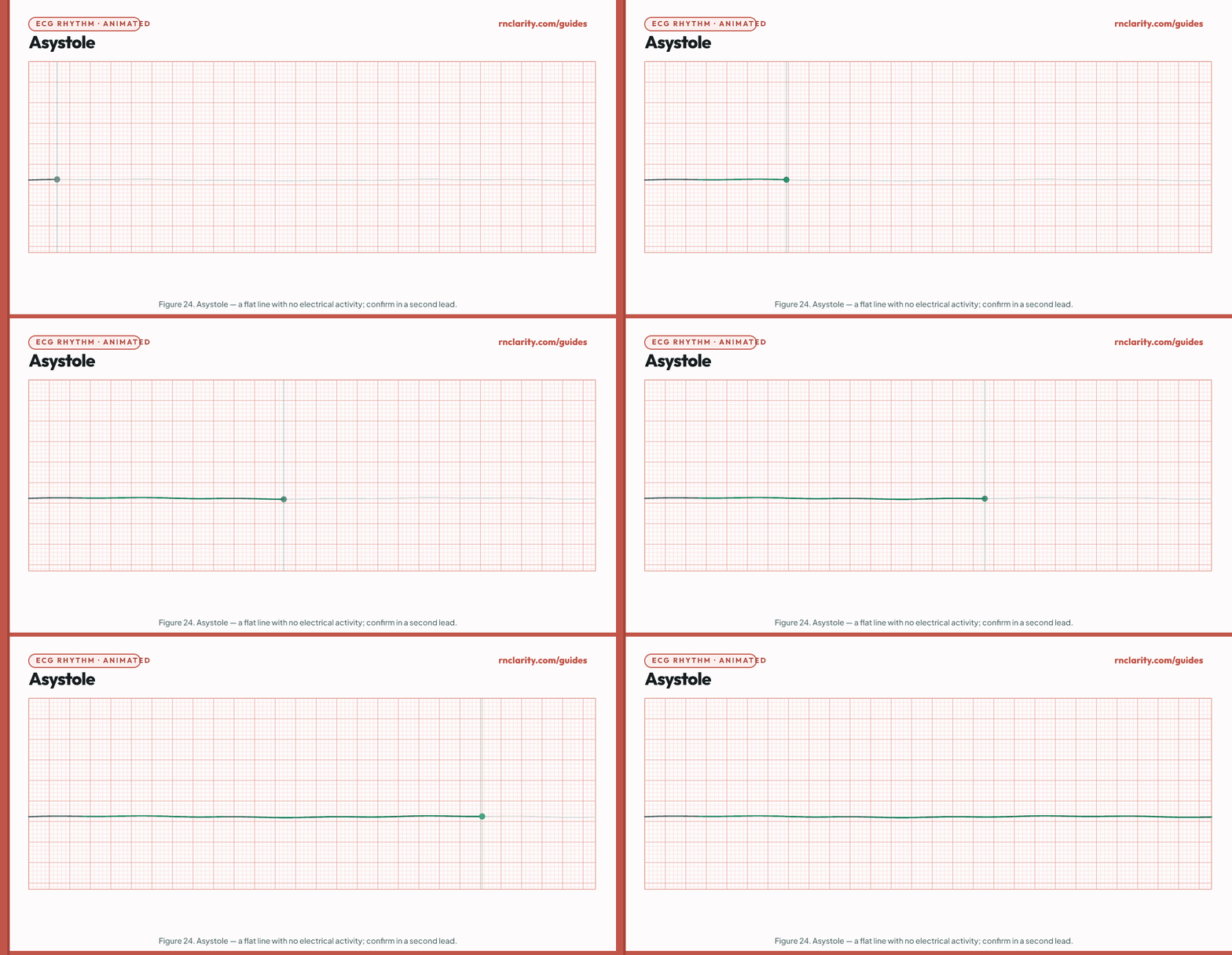
Asystole is the absence of any cardiac electrical activity — a flat line on the ECG. It represents the complete cessation of all electrical impulses. The nurse must confirm asystole in at least two leads to rule out artifact (a single-lead flat line may be a detached lead or artifact).
Nursing priority: Asystole is treated with high-quality CPR and epinephrine 1 mg IV/IO every 3–5 minutes. Defibrillation is not indicated for asystole. The nurse must identify and treat reversible causes (the Hs and Ts): Hypoxia, Hypovolemia, Hydrogen ion (acidosis), Hypo/Hyperkalemia, Hypothermia; Tension pneumothorax, Tamponade, Toxins, Thrombosis (pulmonary or coronary). A new onset of asystole carries a very poor prognosis unless a reversible cause is found and corrected rapidly.
NCLEX-RN® trap: You cannot defibrillate asystole. Defibrillation is only indicated for pulseless VT and VF — shockable rhythms.
13.3 Pulseless electrical activity
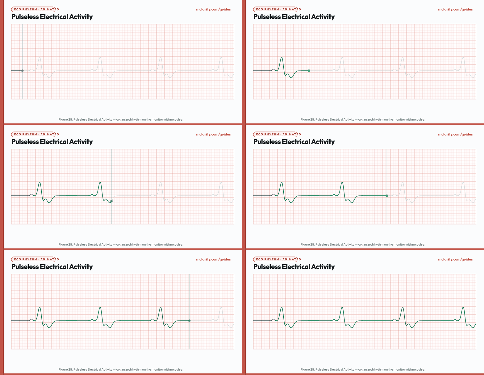
Pulseless electrical activity (PEA) is defined by organized electrical activity on the monitor with the complete absence of a palpable pulse. The ECG may show what appears to be a sinus rhythm, junctional rhythm, or idioventricular rhythm — but the heart's mechanical contraction is not producing output.
Nursing priority: PEA is not defibrillated (the rhythm appears organized). Treatment is high-quality CPR, epinephrine 1 mg IV/IO every 3–5 minutes, and aggressive search for reversible causes (the same Hs and Ts as asystole). The most common reversible causes of PEA include severe hypovolemia (e.g., massive hemorrhage), tension pneumothorax, cardiac tamponade, and massive pulmonary embolism. Identifying and treating the cause within minutes is the only path to survival.
14. Paced Rhythms and Artifact
14.1 Paced rhythm
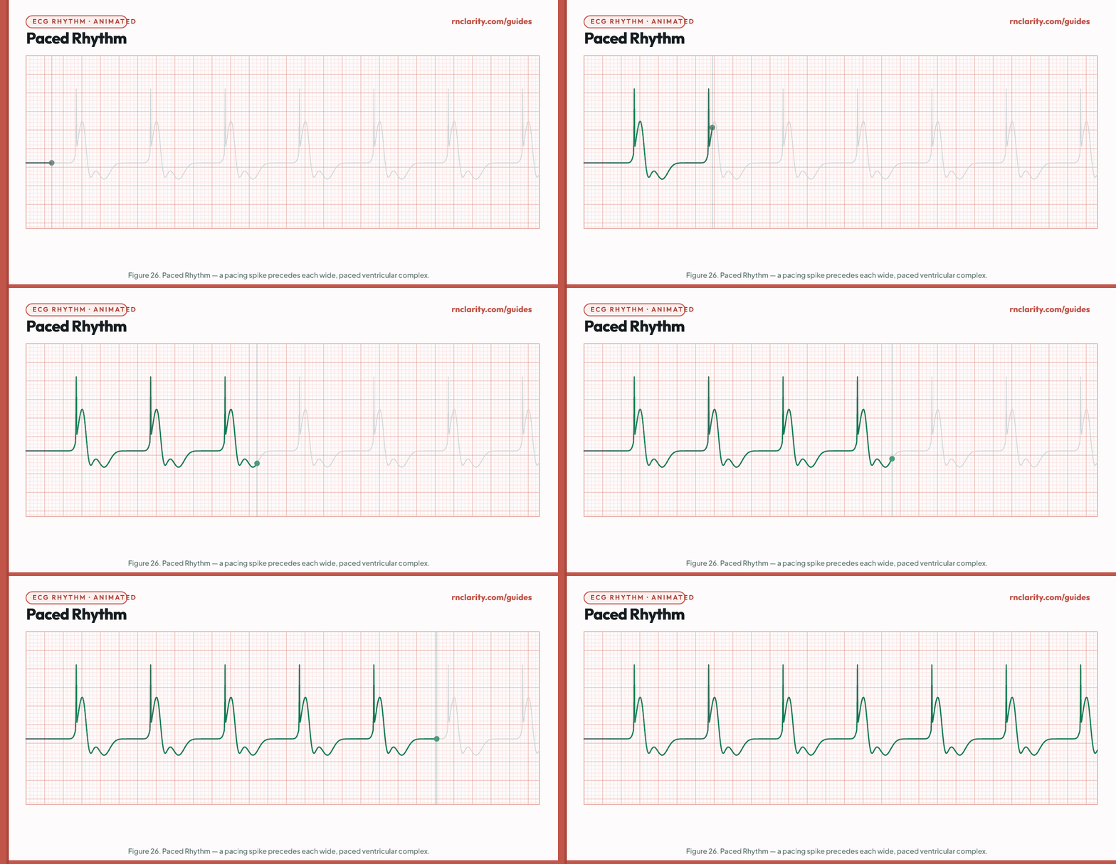
A paced rhythm is identified by the presence of pacemaker spikes — narrow vertical lines immediately before the paced waveform. Atrial pacing produces a spike before the P wave; ventricular pacing produces a spike before a wide, bizarre QRS complex (because ventricular conduction is cell-to-cell, not through the His-Purkinje system).
| Feature | Paced rhythm finding |
|---|---|
| Pacemaker spike | Vertical line immediately before the paced waveform |
| Paced P wave | Small deflection after atrial pacing spike; may not always be visible |
| Paced QRS | Wide (> 0.12 sec), left bundle branch block–type morphology for right ventricular pacing |
| Rate | Programmed rate (commonly 60–70/min in demand mode) |
| Capture | Every spike should produce a P wave (atrial) or QRS (ventricular); failure to capture = spike not followed by waveform |
| Sensing | Pacemaker detects intrinsic beats and withholds firing; failure to sense = pacemaker fires inappropriately (competes with intrinsic beats) |
Nursing priority: Assess for pacemaker function at every shift. Identify the pacemaker type, programmed rate, and mode from the client's chart. Report: failure to capture (spike not followed by waveform), failure to sense (pacemaker fires when the client's own rate is above the set rate), pacing at a rate significantly different from the programmed rate. Assess for pacemaker syndrome (fatigue, dizziness, palpitations due to loss of AV synchrony with single-chamber VVI pacing).
14.2 Motion artifact
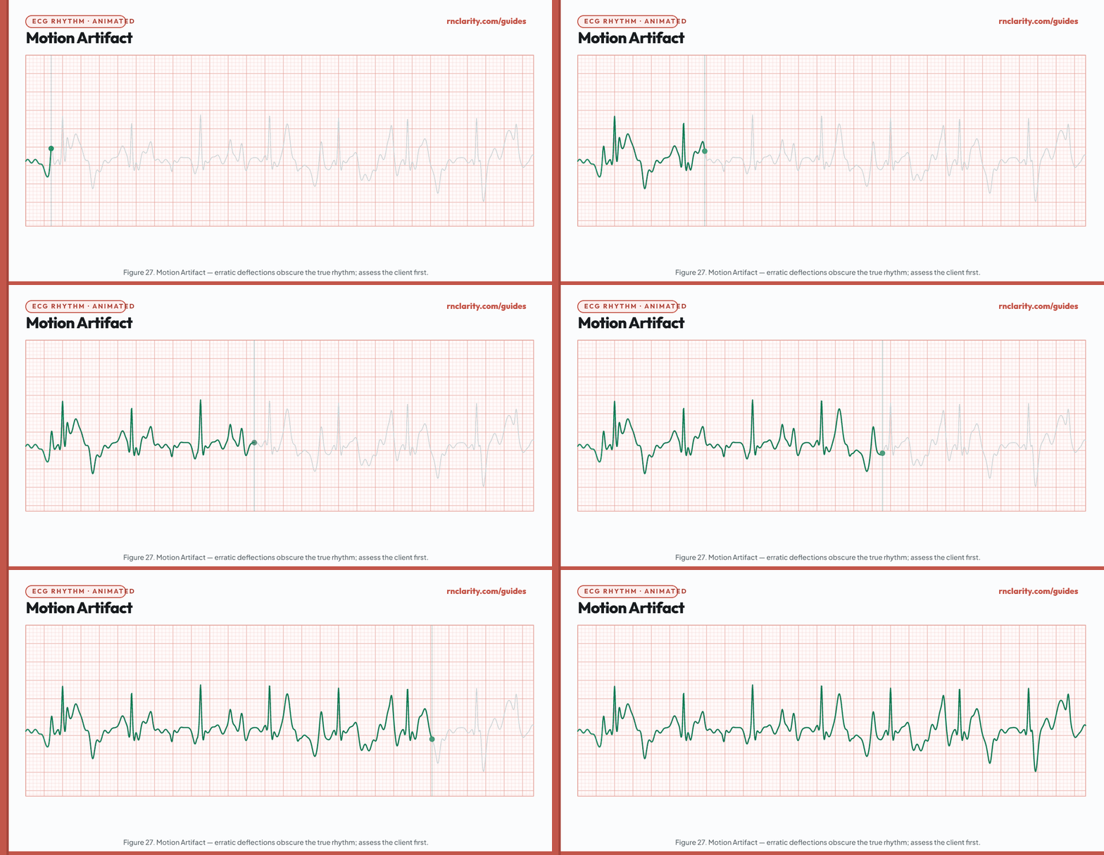
Motion artifact is caused by client movement, shivering, patient transport, or lead wire interference. It appears as erratic, high-amplitude deflections that obscure the true ECG and can mimic VF or other dangerous rhythms. The nurse must always assess the client before treating the monitor.
Clinical rule: If the client is awake, talking, and has a palpable pulse with a normal blood pressure, the monitor is showing artifact — not cardiac arrest. Apply the clinical rule: treat the client, not the tracing.
Troubleshooting artifact:
- Verify all lead connections and ensure electrodes are firmly attached with dry skin beneath.
- Replace aged or diaphoresis-soaked electrodes.
- Ask the client to hold still if possible.
- Use a fresh electrode site and abrade the skin lightly before application.
- Consider a different monitoring lead if one lead shows persistent artifact.
15. Priority Nursing Actions for Bradycardia and Tachycardia
Two broad clinical scenarios — symptomatic bradycardia and unstable tachycardia — trigger the most urgent ECG-related nursing responses. The NCLEX-RN® frequently tests whether candidates can correctly prioritize these actions.
15.1 Bradycardia with cardiopulmonary compromise
Signs of cardiopulmonary compromise: Hypotension, acutely altered mental status, signs of shock, ischemic chest discomfort, or acute heart failure. Assess the client and rhythm together; a slow heart rate alone does not establish the need for atropine.
Priority nursing actions in sequence:
- Stay with the client; call for immediate assistance.
- Maintain the airway, support breathing and oxygenation as indicated, apply the cardiac monitor, and ensure IV access.
- Notify the provider and activate the rapid-response team.
- Prepare atropine 1 mg IV bolus per the adult bradycardia algorithm; administer per order or protocol.
- If atropine is ineffective, prepare transcutaneous pacing and/or a dopamine or epinephrine infusion per the resuscitation protocol; identify and treat the underlying cause.
- Continuously reassess blood pressure, level of consciousness, and SpO₂.
- Document rhythm strip, interventions, and client response.
Adult atropine dose: 1 mg IV bolus; repeat every 3–5 minutes if needed, to a maximum total of 3 mg. See the 2025 AHA adult bradycardia algorithm. This is the adult bradycardia dose; other indications and pediatric care use different dosing.
15.2 Unstable tachycardia (rate > 100/min with hemodynamic compromise)
Symptoms that indicate instability: Hypotension, acute chest pain, signs of heart failure (dyspnea, S3, rales), altered mental status, signs of poor perfusion.
Priority nursing actions in sequence:
- Assess the client; call for immediate assistance.
- Apply oxygen; ensure IV access; attach defibrillator/monitor.
- Notify the provider and prepare for synchronized cardioversion (sedation if time permits).
- For VT with pulse, unstable: synchronized cardioversion 100–200 J biphasic (per ACLS).
- For pulseless VT or VF: unsynchronized defibrillation (200 J biphasic) + CPR.
- Document strip, interventions, cardioversion energy, and response.
Synchronized vs. unsynchronized cardioversion:
| Synchronized | Unsynchronized (defibrillation) | |
|---|---|---|
| Timing | Synchronized to R wave to avoid R-on-T | Not synchronized |
| Indications | Unstable SVT, unstable AF, unstable Aflutter, pulsed VT | VF, pulseless VT |
| Energy (biphasic) | 100–200 J typical (SVT 50–100 J) | 200 J |
| Sedation | Provided if client is conscious | Not needed (client is unconscious) |
Go deeper: For how the NCLEX-RN® tests clinical judgment in cardiac emergencies, see the clinical judgment guide.
16. Electrolytes, Medications and Reversible Causes
ECG changes are often the first clinical sign of electrolyte imbalance or medication toxicity. Recognizing these patterns on the NCLEX-RN® requires connecting the rhythm to the cause.
16.1 Electrolyte effects on the ECG
| Electrolyte disturbance | Characteristic ECG changes | Nursing priority |
|---|---|---|
| Hypokalemia (K⁺ < 3.5 mEq/L) | Flattened or inverted T waves; prominent U waves; prolonged QU interval; ST depression; increased PVC frequency | Replace potassium; check magnesium; monitor for torsades; hold digoxin |
| Hyperkalemia (K⁺ > 5.0 mEq/L) | May show tall, narrow T waves, PR prolongation, QRS widening, or other changes; a normal-looking ECG does not exclude danger and findings need not follow a fixed sequence | For concerning ECG changes, activate the emergency pathway: IV calcium stabilizes the myocardium; insulin with glucose shifts potassium; arrange potassium removal according to renal function and local protocol. Bicarbonate is context-dependent. |
| Hypomagnesemia (Mg²⁺ < 1.5 mEq/L) | Prolonged QT; torsades de pointes; increased PVC frequency; refractory hypokalemia | Magnesium sulfate IV; correct potassium simultaneously |
| Hypermagnesemia (Mg²⁺ > 2.5 mEq/L) | Prolonged PR; wide QRS; flattened T; cardiac arrest at very high levels | Calcium gluconate (antidote); stop magnesium infusion; may require dialysis |
| Hypocalcemia (Ca²⁺ < 8.5 mg/dL) | Prolonged QT (ST segment lengthened); torsades risk | IV calcium gluconate; correct magnesium |
| Hypercalcemia (Ca²⁺ > 10.5 mg/dL) | Shortened QT; short ST segment | IV fluids; calcitonin; bisphosphonates; dialysis |
Critical NCLEX-RN® pattern: Suspect hyperkalemia when a patient at risk has new tall T waves, conduction delay, or QRS widening, but do not wait for a textbook sequence or rule it out because the ECG lacks peaked T waves. Obtain a potassium result, place the patient on cardiac monitoring, and escalate urgently when the clinical picture or ECG is concerning. Sodium polystyrene sulfonate (Kayexalate) is not emergency therapy for life-threatening hyperkalemia because its onset is delayed, as stated in its FDA label.
Go deeper: Acid-base disturbances — especially metabolic acidosis — directly shift potassium out of cells and drive the electrolyte-induced ECG changes described above. For the full four-step ABG interpretation method and how pH, PaCO₂ and HCO₃⁻ connect to dysrhythmia risk, see the ABG interpretation guide.
16.2 Medication-induced ECG changes
| Medication | ECG effect | Nursing concern |
|---|---|---|
| Digoxin (therapeutic) | "Scooped" ST depression; shortened QT; slow heart rate | Document baseline ST morphology; "dig dip" is not ischemia |
| Digoxin toxicity | Bradycardia; any dysrhythmia; AV blocks; accelerated junctional rhythm; PAT with block | Hold digoxin; check serum level (therapeutic 0.5–2 ng/mL); check potassium; Digibind if severe |
| Beta-blockers | Sinus bradycardia; prolonged PR; AV block | Monitor heart rate; assess for symptomatic bradycardia |
| Calcium channel blockers (non-DHP) | Sinus bradycardia; prolonged PR; AV block | Same as beta-blockers; calcium gluconate is the antidote for toxicity |
| Tricyclic antidepressants (TCAs) | Wide QRS; prolonged QT; right bundle branch block; terminal R wave in aVR in overdose | Sodium bicarbonate for wide QRS in TCA overdose |
| Class Ia antiarrhythmics (quinidine, procainamide) | Prolonged QT; torsades risk | Monitor QTc before and during therapy |
| Class III antiarrhythmics (amiodarone, sotalol) | Prolonged QT; bradycardia | Monitor QTc; thyroid function (amiodarone) |
| Atropine | Increased heart rate; shortens PR interval | Monitor for tachycardia; urinary retention in older adults |
16.3 The Hs and Ts of reversible cardiac arrest causes
The 2020 AHA ACLS guidelines identify ten reversible causes of pulseless cardiac arrest organized as the Hs and Ts:
| H causes | T causes |
|---|---|
| Hypovolemia | Tension pneumothorax |
| Hypoxia | Tamponade (cardiac) |
| Hydrogen ion (acidosis) | Toxins |
| Hypo/Hyperkalemia | Thrombosis — pulmonary |
| Hypothermia | Thrombosis — coronary (MI) |
For each arrest scenario on the NCLEX-RN®, consider whether one of these reversible causes is present and can be corrected to improve survival.
Go deeper: For pharmacological interventions used in cardiac emergencies and toxicology, see the pharmacology guide.
17. Clinical-Judgment Cases
The following two cases apply the CJMM six-step framework to ECG interpretation scenarios typical of NCLEX-RN® NGN questions.
Case 1: Newly detected irregular rhythm in a post-surgical client
Clinical vignette: A 68-year-old client is on day 2 following a right total hip replacement. Vital signs are: BP 110/72, HR 118 with irregular pulse, RR 20, SpO₂ 94% on 2 L nasal cannula, temperature 37.8°C. The telemetry monitor shows an irregularly irregular rhythm with no identifiable P waves and narrow QRS complexes. The client reports mild fatigue and "feeling like my heart is skipping."
Step 1 — Recognize cues: Irregularly irregular rhythm, absent P waves, narrow QRS, rate 118/min, SpO₂ 94%, post-surgical state (PE risk), fatigue, mild symptoms of inadequate perfusion.
Step 2 — Analyze cues: The rhythm is consistent with atrial fibrillation with a rapid ventricular rate. Post-surgical AF is common after orthopedic procedures. SpO₂ of 94% suggests mild hypoxia, which can trigger AF. The combination of AF + SpO₂ 94% + post-surgical state also raises concern for pulmonary embolism.
Step 3 — Prioritize hypotheses: (1) New-onset atrial fibrillation with rapid ventricular response — most likely; (2) Pulmonary embolism — must be considered given clinical context.
Step 4 — Generate solutions: Increase oxygen to improve SpO₂ to ≥ 95%; notify the provider immediately with SBAR communication; obtain a 12-lead ECG; prepare for possible rate-control therapy; assess for anticoagulation need; consider Wells PE score if symptoms develop.
Step 5 — Take actions: Apply oxygen; document rhythm strip; notify provider with vital signs, rhythm description, and current symptoms; ensure IV access is patent; gather documentation for anticoagulation eligibility.
Step 6 — Evaluate outcomes: Reassess heart rate, blood pressure, SpO₂, and level of consciousness every 5–15 minutes. Expect heart rate to decrease to 60–100/min with rate-control therapy. Escalate if the client becomes hemodynamically unstable.
Case 2: Client with chest pain and new ECG findings
Clinical vignette: A 72-year-old client with hypertension and type 2 diabetes reports sudden onset of severe chest pain rated 8/10 with radiation to the left jaw for 20 minutes. Vital signs: BP 158/94, HR 52 and regular, RR 18, SpO₂ 97%. The telemetry strip shows a sinus rhythm with a PR interval of 0.24 seconds and ST elevation in leads II, III, and aVF.
Step 1 — Recognize cues: New ST elevation (inferior leads), chest pain with jaw radiation, first-degree AV block (new), bradycardia, hypertension, diabetes, 20-minute duration.
Step 2 — Analyze cues: ST elevation in inferior leads (II, III, aVF) is consistent with inferior STEMI (right coronary artery territory). The right coronary artery also supplies the AV node, explaining the first-degree AV block. Bradycardia accompanies inferior MI due to increased vagal tone and RCA ischemia. This is a time-sensitive emergency.
Step 3 — Prioritize hypotheses: Inferior STEMI with first-degree AV block and vagally mediated bradycardia — highest priority.
Step 4 — Generate solutions: Activate the catheterization laboratory team or STEMI protocol; administer aspirin 325 mg (unless contraindicated); obtain 12-lead ECG; prepare for right-sided ECG leads (V4R) to detect right ventricular involvement; establish two large-bore IVs; monitor for progression of AV block; avoid nitroglycerin if right ventricular MI is present (preload-dependent).
Step 5 — Take actions: Notify provider/cardiologist; activate STEMI protocol; administer medications per protocol; obtain additional ECG leads; prepare transfer to catheterization laboratory.
Step 6 — Evaluate outcomes: Assess for return to sinus rhythm and resolution of ST changes after reperfusion. Monitor for progression from first-degree to higher-degree AV block (Mobitz I, Mobitz II, or complete block) — common with inferior MI. Prepare transcutaneous pacing on standby.
Go deeper: For practice with full six-item NGN cases that apply these steps, see the NGN case studies guide.
18. Practice Questions With Answers and Rationales
The following ten original questions are aligned to the April 2026 NCLEX-RN® Test Plan.
Question 1
A nurse is monitoring a client in the telemetry unit who had a cardiac catheterization six hours ago. The monitor shows a regular rhythm at 42 beats per minute with upright P waves and narrow QRS complexes. The client is alert, blood pressure is 102/64 mmHg, and reports mild dizziness when standing. Which action should the nurse take first?
A) Administer atropine 1 mg IV
B) Assess the client's apical pulse and blood pressure in the supine position
C) Increase the IV fluid rate to 125 mL/hr
D) Place the client in Trendelenburg position
Correct answer: B
Rationale: The rhythm is sinus bradycardia at 42/min. While symptomatic, the nurse's first action is to complete a focused assessment (apical pulse and blood pressure supine) to quantify hemodynamic status before initiating any intervention. Assessment always precedes action on the NCLEX-RN® unless the scenario describes an immediate life threat. Option A (atropine) may be correct later, but the nurse must first establish a baseline and notify the provider. Option C is not ordered. Option D is not appropriate for bradycardia.
Question 2
A nurse reviews a rhythm strip and identifies: rate 78/min, regular rhythm, upright P waves, PR interval 0.28 seconds, QRS 0.08 seconds. Which dysrhythmia does the nurse identify?
A) Normal sinus rhythm
B) First-degree AV block
C) Second-degree AV block type I
D) Second-degree AV block type II
Correct answer: B
Rationale: A PR interval of 0.28 seconds exceeds the upper limit of normal (0.20 seconds). Every P wave is still followed by a QRS complex (one-to-one conduction is maintained) and the PR interval is consistent — this is first-degree AV block, not a higher degree. Second-degree blocks would have P waves not followed by QRS complexes (dropped beats). Normal sinus rhythm has a normal PR.
Question 3
A client is found unresponsive. CPR is in progress. The cardiac monitor shows a completely flat isoelectric line in leads I and II. Which action should the nurse prepare for first?
A) Defibrillation at 200 J biphasic
B) Synchronized cardioversion
C) Confirm asystole in a second lead and assess lead connections
D) Administer amiodarone 300 mg IV bolus
Correct answer: C
Rationale: A flat line may represent true asystole or may be artifact from a loose lead. Before calling asystole and directing interventions, the nurse confirms the rhythm in at least two leads and checks lead connections. True asystole is not treated with defibrillation (option A) — defibrillation is for VF and pulseless VT. Synchronized cardioversion (B) is for organized rhythms with pulse. Amiodarone (D) is indicated for VF/pulseless VT after the first shock, not for asystole.
Question 4
A client receiving IV magnesium sulfate for eclampsia prevention develops bradycardia at 44 beats per minute and the deep tendon reflexes are absent. Which action should the nurse take first?
A) Administer atropine 1 mg IV
B) Stop the magnesium infusion and administer calcium gluconate IV
C) Increase the rate of the magnesium infusion
D) Notify the provider and document the findings
Correct answer: B
Rationale: Absent deep tendon reflexes are the first sign of magnesium toxicity; bradycardia confirms cardiovascular effects. The nurse must stop the magnesium infusion immediately and administer calcium gluconate, the antidote for magnesium toxicity. This is a priority intervention, not merely a notification (option D, which should follow but not precede treatment). Option A does not address the cause. Option C would worsen toxicity.
Question 5
A client with heart failure is receiving digoxin 0.125 mg PO daily. The morning serum potassium is 2.8 mEq/L. Which nursing action has the highest priority?
A) Administer the morning dose of digoxin as prescribed
B) Hold the digoxin, notify the provider, and report the potassium level
C) Administer IV potassium 40 mEq as a rapid IV push
D) Obtain a stat ECG and continue with the digoxin dose
Correct answer: B
Rationale: Hypokalemia (K⁺ < 3.5 mEq/L) dramatically increases the risk of digoxin toxicity. The nurse should hold the digoxin and notify the provider before administration. Option C contains a critical safety error — IV potassium is never given as an IV push; it must be diluted and infused with cardiac monitoring. Option A is unsafe. Option D partially addresses monitoring but does not address the medication safety issue.
Question 6
The nurse is monitoring a client and the telemetry strip shows: rate 186/min, regular, no discernible P waves, narrow QRS complexes (0.08 sec). The client is alert, blood pressure is 118/76 mmHg, and denies chest pain. Which intervention is most appropriate?
A) Prepare for immediate synchronized cardioversion
B) Initiate CPR
C) Assist the client to perform the Valsalva maneuver
D) Administer amiodarone 300 mg IV bolus
Correct answer: C
Rationale: The rhythm is consistent with supraventricular tachycardia (SVT). The client is stable (alert, BP 118/76, no chest pain), so the first-line intervention for stable SVT is vagal maneuvers such as the Valsalva maneuver. Option A (cardioversion) is reserved for unstable SVT. Option B (CPR) is not indicated for a client with a pulse. Option D (amiodarone bolus) is for VF/pulseless VT and refractory VT.
Question 7
A client's telemetry strip shows a PR interval that progressively lengthens over three consecutive beats, followed by a P wave with no QRS, then the cycle resets. Which dysrhythmia does the nurse identify?
A) First-degree AV block
B) Second-degree AV block type I (Wenckebach)
C) Second-degree AV block type II (Mobitz II)
D) Third-degree (complete) AV block
Correct answer: B
Rationale: The pattern of progressively lengthening PR intervals followed by a dropped QRS, then reset, is the hallmark of second-degree AV block type I (Wenckebach / Mobitz I). In first-degree block (A), the PR is prolonged but constant and no QRS is ever dropped. In Mobitz II (C), the PR is constant and the QRS drops without warning. In third-degree block (D), there is no consistent relationship between P waves and QRS complexes.
Question 8
A nurse responds to a cardiac monitor alarm. The client's monitor shows a chaotic, irregular waveform with no recognizable P waves, QRS complexes, or T waves. The client is unresponsive with no pulse. Which action should the nurse take first?
A) Notify the provider
B) Call for the crash cart and defibrillator
C) Initiate chest compressions and call for assistance
D) Administer epinephrine 1 mg IV
Correct answer: C
Rationale: The rhythm is ventricular fibrillation (VF) and the client is in cardiac arrest. The first action per BLS and ACLS is to call for help and begin high-quality CPR while a defibrillator is obtained. Defibrillation is the definitive treatment for VF, but CPR must begin immediately — not delayed to retrieve equipment. Option B is the next simultaneous step. Option D (epinephrine) comes after the initial defibrillation attempt. Option A (notify provider) is not the first priority in a cardiac arrest.
Question 9
A nurse is caring for a client on day 3 following an anterior wall myocardial infarction. The telemetry strip shows a regular rhythm at 44 beats per minute. P waves are visible at a rate of 78/min, but they have no consistent relationship to the QRS complexes, which are wide at 0.16 seconds. Which interpretation is correct?
A) Second-degree AV block type II with bundle branch block
B) Third-degree (complete) AV block with ventricular escape rhythm
C) Junctional escape rhythm with aberrant conduction
D) Sinus bradycardia with bundle branch block
Correct answer: B
Rationale: The presence of two independent rates (P waves at 78/min, QRS at 44/min) with no consistent PR interval is diagnostic of third-degree (complete) AV block. The wide QRS (0.16 sec) indicates a ventricular escape pacemaker. In anterior MI, complete AV block results from extensive damage to the bundle branches. This rhythm requires emergency notification and preparation for transcutaneous pacing. Options A, C, and D all describe rhythms with some relationship between P waves and QRS.
Question 10
Which electrolyte disturbance should the nurse suspect when a client's ECG shows tall, narrow, symmetrically peaked T waves in multiple leads?
A) Hypokalemia
B) Hyperkalemia
C) Hypomagnesemia
D) Hypercalcemia
Correct answer: B
Rationale: Tall, narrow, symmetric (tent-shaped) peaked T waves are the earliest and most characteristic ECG sign of hyperkalemia. This finding appears when the serum potassium exceeds approximately 5.5–6.0 mEq/L. Hypokalemia (A) produces flattened T waves and prominent U waves. Hypomagnesemia (C) prolongs the QT interval. Hypercalcemia (D) shortens the QT interval. Recognizing peaked T waves in a client with renal failure, acidosis, or on ACE inhibitors/potassium-sparing diuretics should prompt an immediate potassium level check.
Go deeper: For additional NCLEX-RN® practice questions with full rationales across all clinical categories, see the practice questions guide.
References
The content of this guide is based on the following publicly available authoritative sources. No copyrighted test bank material is reproduced.
-
National Council of State Boards of Nursing (NCSBN). 2026 NCLEX-RN® Examination: Test Plan for the National Council Licensure Examination for Registered Nurses. Effective April 1, 2026. Chicago: NCSBN; 2025. Available from: ncsbn.org.
-
American Heart Association. Adult Bradycardia With a Pulse Algorithm (2025). Current adult atropine dose and escalation pathway.
-
Dubin D. Rapid Interpretation of EKG's. 6th ed. Fort Lauderdale: Cover Publishing Company; 2000.
-
Phalen T, Aehlert BJ. The 12-Lead ECG in Acute Coronary Syndromes. 3rd ed. St. Louis: Elsevier; 2012.
-
Jacobson C. AACN Advanced Critical Care. Chapters on dysrhythmia interpretation. Published by the American Association of Critical-Care Nurses.
-
Goldberger AL, Goldberger ZD, Shvilkin A. Goldberger's Clinical Electrocardiography: A Simplified Approach. 9th ed. Philadelphia: Elsevier; 2018.
-
Drew BJ, et al. "Practice Standards for Electrocardiographic Monitoring in Hospital Settings." Circulation. 2004;110(17):2721–2746. American Heart Association Scientific Statement.
-
American Heart Association. Part 9: Adult Advanced Life Support (2025).
-
Zipes DP, et al. Braunwald's Heart Disease: A Textbook of Cardiovascular Medicine. 11th ed. Philadelphia: Elsevier; 2019.